- Table View
- List View
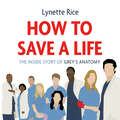
How to Save a Life: The Inside Story of Grey's Anatomy
by Lynette RiceThe first inside story of one of TV's most popular and beloved dramas, Grey's Anatomy.More than 15 years after its premiere, Grey's Anatomy remains one of the most beloved dramas on television in the US and the UK. It continues to win its time slot and has ranked in the Top 20 most watched shows in primetime for most of its 17-season run. It currently averages more than 9 million viewers each week. More than that, it's been a cultural touchstone. It introduced the unique voice and vision of Shonda Rhimes, it made Ellen Pompeo, Sandra Oh and T.R. Knight household names, and injected words and phrases into the cultural lexicon like 'McDreamy,' and 'you're my person.' And the behind-the-scenes drama has always been just as juicy as what was happening in front of the camera, from the high-profile firing of Isaiah Washington to Katherine Heigl's fall from grace and Patrick Dempsey's shocking death episode. The show continued to haemorrhage key players, but the beloved hospital series never skipped a beat. Lynette Rice's How to Save A Life takes a totally unauthorized deep dive into the show's humble start, while offering exclusive intel on the behind-the-scenes culture, the most heartbreaking departures and the more polarizing plotlines. It's the perfect listen for all Grey's Anatomy stans out there.(P) 2021 Blackstone Publishing
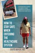
How to Stay Safe When Entering the Healthcare System: A Physician Walks across the Country to Raise Awareness of the Need to Improve Healthcare Safety
by David MayerThis book is an urgent call to action centering on the author's thirty-five-year mission to raise awareness of the 250,000 lives that are lost each year to preventable medical harm and the harm faced by healthcare professionals in the form of workplace vi
How to Succeed as a Leader (How To Succeed Ser.)
by Richard Jones Kay Mohanna David Wall Ruth ChambersThis work includes Foreword by David Nicholson - Chief Executive, National Health Service of England. In the past, there has been too little emphasis and investment made in developing leaders in healthcare. People have become leaders without being prepared or trained or supported in the role. Individuals need to understand the context, the concept and models of good leadership, the practical steps to becoming a good leader, and how to sustain the various components of a well functioning and effective organisation, whether that is a large NHS trust or hospital department, a clinical group or practice team. This guide has been written by a range of writers from organisational consultancy and NHS backgrounds who are all experienced in developing and supporting leaders, planning and providing education, and change management. It is specially designed for independent learning, with answers to frequently asked questions, self-assessment exercises and helpful tips. "How to Succeed as a Leader" is ideal for all healthcare professionals in (or aspiring to) leadership roles. It also provides inspiration for academics and workplace educators, managers and leaders in government, strategic health authorities and workforce deaneries. 'There is constant reorganisation and a changing culture in our health service. Good leadership is essential to address the changes required and take others with you so that the service can function effectively. There has been an amateurish approach to leadership in the NHS in the past, where people have become leaders without being prepared or trained for the role or supported in it. This book is all about presenting you with a practical approach to becoming a competent leader, to prepare you to lead in a positive way and realise your responsibilities as a leader.' From the Preface.
How to Succeed at E-learning
by Paul Kirk Peter Donnelly Joel BensonA basic guide to getting the best from e-learning for medical students, teachers and all healthcare professionalsHow to Succeed at e-Learning answers the needs of all healthcare professionals either starting or continuing their studies but not knowing where to begin with e-learning. It is a valuable guide for learners in undergraduate and postgraduate medicine as well as related health professionals and essential for teachers of medicine who are beginning to transfer from print to electronic teaching and need to understand effective methods of presentation.
How to Succeed at Interprofessional Education (How To)
by Peter DonnellyHealthcare professionals work not only within multidisciplinary teams but also with allied health and non-healthcare professionals. How to Succeed at Interprofessional Education is a practical introduction to the concepts of interprofessional education (IPE) within medicine. It provides the rationale and framework for the effective delivery of IPE in a range of health care contexts. The contents include definitions, the evidence that IPE is effective, and the principles to ensure successful delivery. A series of examples of IPE in different clinical settings is described that lead to improved decision making and improved clinical care for patients.
How to Succeed at Medical School
by Dason Evans Jo BrownCan you adapt to the wide variety of learning environments in medicine? Can you show your best abilities in the exams at the same time as learning to be a doctor? Can you balance your studies with an enjoyable social life? Can you develop your professionalism and manage your 'digital footprint'? How to Succeed at Medical School will help you learn these vital skills, and much more. Written by experienced medical school teachers and packed full of case studies, illustrations, quotes from other students, tip boxes, exercises, portfolios and learning techniques to help you communicate, study and revise - it's an essential resource to help you thrive at medical school. This thoroughly updated second edition includes new chapters on Professionalism and Teaching, and provides invaluable insight into what to expect from the start of medical school right through to the start of your medical career.
How to Succeed at Medical School: An Essential Guide to Learning (How To #25)
by Dason Evans Jo BrownCan you adapt to the wide variety of learning environments in medicine? Can you learn for exams at the same time as training to be a doctor? Can you stay focused on the future while getting today’s job done? Can you achieve a life-work balance? How to Succeed at Medical School will help you learn these vital skills, and much more. This excellent guide to the study skills essential for surviving and thriving at medical school gives you insight into what to expect, covering the early days right through to clinical attachments. With case studies, illustrations, quotes from other students, tip boxes, exercises, portfolios, and learning techniques to help you communicate and to study and revise — it’s jam-packed to help you succeed! Written by experienced medical school teachers, this is your guide from the start of medical school to the start of your medical career. Pre-publication reviews: "… I learned a lot, found the enthusiasm of the text motivating and inspiring and really enjoyed reading it." –Second year medical student, Royal Free and UCL "I just wish this book had been available when I started my clinical placements." –Second year medical student, University of Liverpool "It helps aid students to learn effectively and efficiently and even tells you how you will know when you know enough!" –Professor Parveen Kumar
How to Succeed at Revalidation (How To)
by Peter Donnelly Katie WebbDoctors in the UK are required to provide evidence of their fitness to practice—all doctors registered with the General Medical Council (GMC) need to revalidate to maintain their registration. How to Succeed at Revalidation contains up-to-date information on the current regulatory framework and step-by-step guidance to the entire revalidation process. Offering expert advice on how to undertake the process from the perspective of the appraisee, the appraiser, and the employer, this practical, quick-reference guide covers each of the Good Medical Practice (GMP) domains: Knowledge, Skills and Performance; Safety and Quality; Communication, Partnership and Team Work; and Maintaining Trust. Throughout the text, numerous examples describe different models of appraisal and reflection, identify activities that map to each domain, and demonstrate how revalidation requirements can be achieved in the course of daily practice. This much-needed guide: Covers regulatory processes in medicine and healthcare from both UK and international perspectives Reviews the background that led to the implementation of revalidation Discusses the new GMP requirements for all doctors in the UK Explores the possible future of revalidation How to Succeed at Revalidation is essential reading for all qualified and trainee doctors, undergraduate and postgraduate learners, tutors and trainers seeking to navigate the revalidation process in the UK.
How to Succeed at the Medical Interview (How To #27)
by Chris Smith Darryl MeekingDoctors are likely to undergo several interviews of different types during their career, and this new title in the popular 'How to' series aims to guide the medical professional through the steps necessary to thoroughly prepare for this competitive process. Contents include: Discussion of the different types of medical interview How to prepare for interview How to optimise your performance Information about common questions and how best to answer them Information about knowledge-based questions currently asked Coverage of questions that test generic skills and how to answer them How to prepare for competency-based assessments and tasks. An ideal companion for all health professionals faced with an interview, How to Succeed at the Medical Interview will assist in building confidence and ensuring that candidates are as thoroughly prepared as possible.
How to Succeed at the Medical Interview (How To #42)
by Chris Smith Darryl MeekingHow to Succeed at the Medical Interview provides candidates with a competitive edge. It reduces the likelihood of unexpected questions or situations and helps improve confidence before and during the medical interview. This new second edition includes updated content on changes to the structure of healthcare and how this affects both the application and interview process. It details the types of questions that will be asked at medical interviews and also provides improved guidance for overseas doctors and healthcare professionals and for those seeking to practice abroad. How to Succeed at the Medical Interview is the ideal guide for Foundation Programme trainees, Specialist Registrars and General Practitioner trainees. It is also valuable for healthcare professionals facing competitive medical interviews at any stage of their career.
How to Succeed in Medical Research: A Practical Guide (How To)
by Robert Foley Robert Maweni Shahram Shirazi Hussein JaafarHow to Succeed in Medical Research is a practical resource for medical students and junior doctors across all specialties. Designed for busy readers seeking to distinguish themselves in a highly competitive environment, this concise yet comprehensive guide provides step-by-step advice on selecting a project, finding a mentor, conducting a study, analysing results, publishing a paper, communicating findings, and much more. Presented in an accessible and conversational style, 14 succinct chapters walk readers through the essential stages of their research journey, from the initial steps to getting involved in research as a medical student, to effectively balancing clinical work, scientific research, and other academic pursuits early in your career as a healthcare professional. The book is packed with real-world case studies and expert tips to help readers apply the content directly in their own studies and careers. Straightforward and easy-to-use, this valuable guide: Covers a variety of clinical research and presentation skills using clear and engaging language Provides detailed guidance on writing a paper, conducting a clinical audit, creating a CV and portfolio, and other key proficiencies Develops writing skills for literature reviews, critical appraisals, and case reports Discusses how to further medical careers through research electives, PhD studies, teaching, and quality improvement projects Offers a range of helpful learning features including objectives, key points, case studies, review questions, and links to references and further readings Includes PowerPoint templates for oral presentations and posters via a companion website How to Succeed in Medical Research: A Practical Guide is an ideal resource for medical students, junior doctors and other early career medical professionals.
How to Succeed in Psychiatry
by Henning Sass Iris Calliess Andrea FiorilloAimed at recently qualified psychiatrists or those looking to qualify soon, How to Succeed in Psychiatry is not a source of clinical information but a survival guide to help you through the first years practising psychiatry. This book covers the topics you won't find in standard textbooks. It deals with daily problems and practical solutions for young psychiatrists. Psychiatric training is less team based than other specialties, so there is less opportunity for learning from colleagues than one would expect: this book helps to fill that gap.The book opens with an overview of psychiatry training, describing the similarities and differences among various countries. Subsequent chapters address the opportunities for research and how to publish the results. Psychotherapy and community psychiatry each merit their own chapter on training. Next, the book guides you through the transition phase into a job, discussing opportunities in both the public and private sectors and considering how to choose the best career for you. It reviews important general considerations, such as ethics, professionalism, leadership and management, how to avoid stress and burn out, and how to liaise with other specialties. The book closes with an account of the role of psychiatry associations and continuing professional development.Written by early career psychiatrists from around the world, this book provides invaluable first-hand experience to all those wishing to embark on a career in this exciting discipline.Practical tips for young psychiatrists starting their careers on the wards or in private practice Advice on the transition phase at the end of training, career choice and job opportunities
How to Succeed in Writing a Book: Contemporary Issues In Practice And Policy, Parts 1&2, Written Examination Revision Guide (How To Succeed Ser.)
by Ruth Chambers Gill Wakley Gilian Nineham Gregory Moxon Michele TophamThis highly practical text is full of interesting tips and words of advice covering all stages in publishing including proposals, selection of authors, writing, editing, finding the right publisher, managing other authors, self discipline, marketing, and finance. "This is a 'how to do it' book for anyone considering writing a book. It helps inexperienced or frustrated authors realise where they may be going wrong. Learn how to write to be understood. Pick up tips from the authors of this book- who have all been in the writing and publishing business for a long time. Although, the book focuses on writing for health and social care, most of the information and guidance about getting published can be transferred to any kind of book or publication." - From the Preface.
How to Succeed in the Academic Clinical Interview
by Wei Keith Tan Rory PiperAcademic clinical posts offer doctors the highly rewarding opportunity to maintain both clinical and research careers, but these opportunities are fiercely competitive. This book provides medical students and doctors-in-training with a complete guide to preparing, applying and interviewing for such posts. Providing guidance on the typical UK academic pathways (including Academic Foundation Programme (AFP), Academic Clinical Fellowship (ACF), and Academic Clinical Lectureship (ACL)), candidates will learn how to choose a programme that suits their needs and experience. They will also get practical tips on how to best showcase their achievements and work portfolio in order to submit the highest quality application. A range of model answers to application and in-person questions are provided, together with a mock interview section demonstrating how to approach tricky questions and interviewers. Prepare for successful academic clinical interviews by following the tips and advice from authors who have excelled at their own interviews.
How to Succeed on Nursing Placements (Transforming Nursing Practice Series)
by Karen ElcockFeel confident and fully prepared on your nursing placements with this invaluable guide to one of the most important aspects of your nursing course. Covering the what, why and how in easily accessible language, this book explores the common challenges faced by nursing students on placement and gives you practical advice on how to overcome them. Written by a team of experienced lecturers and nurses, the book covers everything from developing resilience to reflecting on your experience and preparing for employment, enabling you to make the most of your time on placement. Key features - Fully mapped to the new NMC standards of proficiency for registered nurses (2018) - Case studies, activities and other learning features help you translate the theory to practice - A practical guide to help you gain the most from your placement
How to Succeed on Primary Care and Community Placements
by David Pearson Sandra NicholsonHow to Succeed on Primary Care and Community Placements offers practical advice on how to get the most from your time on community visits, within patient consultations, and with the practice team. It highlights the unique opportunities and challenges you will face on placement, from using clinical information systems, to home visits and long term patient relationships, and how to take advantage of new ways of learning with web-based tools, mobile devices and social networking.Key features include:* Learning outcomes at the start of each chapter with links to web-based learning, case examples, and tasks to undertake whilst on placement* An evidence-based, practical approach to improving learning, teaching, assessment and feedback in community settingsWritten by a team of experienced community-based medical education specialists, it is ideal for all medical students, whether on early clinical placements or later in training, and for tutors and preceptors looking for novel ways to engage their students.
How to Support the Neuropsychological Health of the Vietnamese Diaspora (A Clinical Guide to the Neuropsychological Health of Immigrant Populations)
by Lauren G. Mai Lindsay N. VoHow to Support the Neuropsychological Health of the Vietnamese Diaspora is the first book in a new series entitled A Clinical Guide to the Neuropsychological Health of Immigrant Populations, which guides clinicians in the art and science of providing culturally competent services to specific communities. Grounded in evidence-based research and clinical experience, the book offers a better understanding of the unique problems and experiences that the Vietnamese population share, along with examples of how to navigate cultural differences in the assessment and treatment of cognitive impairment.The book reviews the sociocultural and historical factors relevant to those of Vietnamese descent, which help to conceptualize individuals' presentations, common socio-cultural considerations for assessment or treatment, and literature related to working with this population in an international and medical context. It also offers current practice guidelines or approaches to assessment and intervention, along with case studies, a glossary of the necessary cognitive science terms in Vietnamese, and practical resources.It is essential reading for clinicians in the patient care setting, as well as students and researchers in clinical neuropsychology and related fields of psychology, sociology, medicine and forensics.
How to Survive Being a Doctor: Tongue-In-Cheek Advice and Cheeky Illustrations about Being a Doctor
by Clive Whichelow Mike HaskinsYour job is rewarding, but if you’re going to be faced with the horrors of the human body you’re going to need survival skills. This mischievous little book will help see you through your years as a doctor with tongue-in-cheek advice and cheeky illustrations.
How to Survive Dental Performance Difficulties (How To (Dentistry))
by Janine BrooksHow to Survive Dental Performance Difficulties offers an authoritative guide for successfully navigating and overcoming dental performance issues. Offers a practical guide for preventing and overcoming dental performance issues Highlights case studies of dental professionals who have direct experience of being referred for fitness to practise issues Includes information on the support available to dental professionals, the requirements that need to be met, and how to meet them Contains information on the effective use of evidence, improvement practice tools such as personal development plans, continuing professional education, reflective diaries, and audits Offers guidance on how to increase self-awareness and insight
How to Survive Losing a Loved One: A Practical Guide to Coping with Your Partner's Terminal Illness and Death, and Building the Next Chapter in Your Life
by Christine Pearson Karen Jackson TaylorA practical, empowering guide to navigating your partner's diagnosis of a terminal or life-limiting illness, or death. Receiving the news that your partner has a terminal or life-limiting illness, or has died unexpectedly, is among the worst experiences in life. At a time when you are least able to cope, you are faced with a multitude of difficult decisions, some of which must be made quickly. What you need is a friend who has experienced everything you are about to face, who can support you as you navigate some tough, important choices. This book is that friend. There is plenty of information out there but where to start looking? What information is needed and how can it be accessed? What decisions are essential in the immediate term and what can be left until later? Throughout the book, the emphasis is on protecting and supporting those left behind by presenting almost every choice you may need to make and the possible implications of each decision. You will learn:- The importance of creating a will, arranging power of attorney, organising advanced decisions of treatment, and even getting married or entering a civil partnership- What you are entitled to from the state, the NHS and your employer- How to stabilise your finances and prepare to run a household alone- Where your partner ought to be during treatment and/or palliative care, and how to go about achieving this- Which decisions need to be made after death, from planning the funeral to accessing your partner's estate- How to navigate the grieving process and take control of a happy future No matter where you are in the process, How to Survive Losing a Loved One is a comprehensive, practical and empowering guide to coping with your partner's terminal illness and death, and building the next chapter in your life.
How to Survive Losing a Loved One: A Practical Guide to Coping with Your Partner’s Terminal Illness and Death, and Building the Next Chapter in Your Life
by Christine Pearson Karen Jackson TaylorA practical, empowering guide to navigating your partner's diagnosis of a terminal or life-limiting illness, or death. Receiving the news that your partner has a terminal or life-limiting illness, or has died unexpectedly, is among the worst experiences in life. At a time when you are least able to cope, you are faced with a multitude of difficult decisions, some of which must be made quickly. What you need is a friend who has experienced everything you are about to face, who can support you as you navigate some tough, important choices. This book is that friend. There is plenty of information out there but where to start looking? What information is needed and how can it be accessed? What decisions are essential in the immediate term and what can be left until later? Throughout the book, the emphasis is on protecting and supporting those left behind by presenting almost every choice you may need to make and the possible implications of each decision. You will learn:- The importance of creating a will, arranging power of attorney, organising advanced decisions of treatment, and even getting married or entering a civil partnership- What you are entitled to from the state, the NHS and your employer- How to stabilise your finances and prepare to run a household alone- Where your partner ought to be during treatment and/or palliative care, and how to go about achieving this- Which decisions need to be made after death, from planning the funeral to accessing your partner's estate- How to navigate the grieving process and take control of a happy future No matter where you are in the process, How to Survive Losing a Loved One is a comprehensive, practical and empowering guide to coping with your partner's terminal illness and death, and building the next chapter in your life.
How to Survive Your Doctor's Care: Get the Right Diagnosis, the Right Treatment, and the Right Experts for You
by Pamela F. GallinDr. Pamela Gallin. She’s one of the worlds leading pediatric surgeons—a world-class physician with an insider’s access to the best health care in the country. And still, an astonishing series of medical goofs and neglect nearly led to permanent nerve damage of her hand. If someone like Dr. Pamela Gallin—who knows the system and maneuvers within it at the most senior levels—can’t get first-class care, what are the chances that you will? Dr. Gallin’s close call with medical disaster opened her eyes to the perils and pitfalls faced by all patients—even savvy off-duty surgeons. Empowered by her unique perspective, Dr. Gallin gives us How to Survive Your Doctor’s Care, the essential insider’s guide for anyone attempting to navigate the health care system. She offers not only a revealing look at today’s complicated medical system but also real-life examples of what can—and did—go wrong. She explains: —How to take advantage of the hidden network of doctors connected to your primary care physician —Why your life may depend on finding the right specialist —When it’s smart to go outside of your insurance plan —How a Magic Marker might be your final defense against a ghastly surgical mishap —Why your choice of physician determines the quality of your anesthesiologist, radiologist, pathologist, and nurses —The secret to getting great care from your nurse—who might just be your most important health care provider and protector —Why seeking second opinions should be second nature Now more than ever, says Dr. Gallin, patients must assume a more active role in their care. How to Survive Your Doctor’s Care offers invaluable counsel on choosing the right hospital; becoming your own best advocate once you get to the hospital; determining which steps to take before you undergo a surgical procedure; and communicating effectively with your doctors and nurses. Inside, you’ll find: y An 11 -point checklist for selecting the hospital that’s right for you / Dramatic, real-life examples of medical sleuthing at its best—and medical neglect at its worst y Step-by-step instructions for turning your cumbersome provider directory into a manageable list of potential primary care physicians y Crucial questions to ask any prospective physician y Guidelines for selecting an advocate to help you manage and keep track of your care Getting quality health care—even safe health care—has never been more of a challenge. But with Dr. Gallin’s guidance and expertise, you’ll have the tools you need to chart a course to recovery and emerge unscathed from the medical care maze.
How to Survive a Medical Malpractice Lawsuit
by Ilene R. BrennerEveryone seeks to avoid getting into a lawsuit, but what do you do if this does happen?Getting sued for medical malpractice is one of the most traumatic events of a physician's career.This text will guide doctors and physicians through the process from the moment they receive a summons until the after-trial appeal process.Containing valuable information that physicians need to know to prevent making critical mistakes that can hurt their caseWith strategies explained to maximize their chances of a defendant's verdict.Including vital information on how to change your attorney, act at the deposition and dress for court,Navigating through what is a mysterious and terrifying process in non-legalese language that is easy to understand including what makes patients angry, strategies for coping, sample questions and tips on answering them to what happens in court and how to continue if there is a bad outcome.