- Table View
- List View
How to be Nowhere
by Tim MacGabhannLife is finally on the right track for reporter and recovering addict Andrew: he is slowly coming to terms with the murder of his photographer boyfriend Carlos, pursuing sobriety and building a new home with a new partner. Andrew has almost forgotten about the story that ruined his life - but that story hasn't forgotten about him, and a series of deadly threats forces him into helping the very man whose gang murdered his boyfriend and left him homeless.A literary take on the classic chase movie, HOW TO BE NOWHERE is the sequel to Tim MacGabhann's genre-busting and critically-acclaimed debut CALL HIM MINE, and a blistering thrill-ride deep into the fog of Central America's murky present and tragic future.
How to be Sick: A Buddhist-Inspired Guide for the Chronically III and their Caregivers
by Toni BernhardThis book will help and inspire those who must live with the challenges posed by any chronic illnesses, and their caregivers: the relentlessness of symptoms; coming to terms with a life of comparative isolation; weathering fear about the future; facing the judgment of others; dealing with the health care system; and, for a spouse, partner, or other caregiver, adapting to so many unexpected and sometimes sudden life changes.
How to be a Good Enough GP: Surviving and Thriving in the New Primary Care Organisations
by Gerhard Wilke Simon FreemanThe upheavals of the NHS reforms have caused a great deal of stress and uncertainty in primary care, and professional development and support for general practitioners needs to take account of this. This book offers a group supervision model which can be used to develop the core competencies needed for GPs to make the new primary care organisations work. The book analyses how primary care professionals have dealt with the various reforms of the past decade, and picks apart the paralysing culture of politeness, conflict avoidance and rivalry for power, to reveal how at the core of reform is the struggle for each GP to construct a new professional identity which integrates medicine, management and politics. It proposes ways GPs can benefit from these experiences to become equipped with the necessary competencies to be active members or dynamic leaders in the new primary care organisations. The doctor-patient relationship is no longer one-to-one, but located within a group matrix, in the same way that a GP is now required to work within a group framework. This book enables GPs to develop the essential group skills they now need, and on which the success of the healthcare reforms ultimately depends.
How to be a Nurse or Midwife Leader
by David Ashton Jamie Ripman Philippa WilliamsHow to be a Nurse or Midwife Leader is an indispensable guide for all nurses and midwives who wish to develop and improve their practice as leaders. Written in collaboration with the NHS Leadership Academy, this practical book draws on the real experience of over 10,000 nurses and midwives to bring leadership dilemmas to life in specific situations. Key learning features include: How to develop your self-awareness How to develop your personal impact and presence How to survive and thrive How to get your message across How to get the best out of others How to work with and lead other professionals and patients How to have courageous conversations How to balance conflicting demands and needs Containing exercises and reflective questions to help apply theory to leadership practice, How to be a Nurse or Midwife Leader is an ideal companion for all nurses and midwives, whether you are newly qualified, or stepping into a team leader role.
How to be a Safe Consultant Vascular Surgeon from Day One: The Unofficial Guide to Passing the FRCS (VASC)
by James Michael ForsythThis is a highly pragmatic and down-to-earth guide to the “real world” of vascular surgery, written to help trainees pass the FRCS (Vasc) Examination the first time around, hopefully with flying colours! Through clear and concise chapters, a review of relevant vascular surgery papers, a solid grounding in up-to-date and relevant UK and European guidelines, and mock examination questions, this book should give candidates a fighting chance at exam success. Using his early experience, and insight gained from senior consultant vascular surgeons and mentors, author James Forsyth dispenses both facts and pearls of wisdom as he takes the prospective vascular surgeon through the core topics pertinent to a day one vascular consultant. This guide specifically covers: • “Building a strong foundation” with a focus on patient counselling, presentation skills, clear documentation, professionalism, and a foundational grasp of the key evidence base for vascular surgery • “Core vascular surgery topics” from chronic limb-threatening ischaemia to aorto-iliac aneurysms to symptomatic carotid artery disease to chronic venous disease • Pragmatic guide to the “bread & butter” vascular surgery operations • Fifty mock SBA assessment questions and answers, and numerous mock FRCS short and long cases that are blended into reality-based discussions to draw out key learning points This book is essential to any budding vascular surgeon intending to sit the FRCS (Vasc) examinations, and those who are about to start off as real day one consultant vascular surgeons. Indeed, the FRCS (Vasc) Examination is a bridge to becoming a true consultant vascular surgeon, and this book therefore is a “must-read” for anyone looking to cross that bridge.
How to use 3D Printing Innovations and Digital Storage to Democratize Anatomy Education (Biomedical Visualization #6)
by Leonard ShapiroThis edited book contains chapters that describe bespoke three-dimensional (3D) printing aimed at democratizing anatomy education by providing open-source scans for download and printing as 3D models. The long history of anatomical models as educational resources is explored in fascinating detail, from wax models through to a range of cutting-edge 3D printers. In a related chapter, a veterinary anatomy educator describes a transformation in teaching and learning methods in veterinary education using Augmented Reality (AR), Virtual Reality (VR) and 3D visualization methods like CT or MRI images which can be used to reconstruct complete 3D virtual models, as well as 3D prints from these reconstructed scans. The first digital, cloud-based human skeletal repository in southern Africa is an extensive and categorized ‘bone library’ globally accessible for use in education and research. A chapter details a digital protocol for the bioprinting of a 3D acellular dermal scaffold (ADS) for use in wound healing, as an alternative to skin grafting for secondary intention wound healing. A chapter offers an extensive guide to applied anatomy for acupuncture and is provided in 4 parts viz, upper limb, lower limb, trunk, head and neck. Each part of the chapter is replete with beautiful cadaveric images including annotations that relate specifically to information in the text. We look at vertebral artery variations and its role in clinical conditions, current insights into polycystic ovarian syndrome, and visual interpretation using multiplex immunoassay of serum samples. This book will appeal to educators of both human and animal anatomy who have a keen interest and focus on the use of bespoke 3D printing, augmented and virtual reality, as well as acupuncture practitioners, clinicians, regenerative medicine specialists, surgeons, tissue engineers and artists.
How's It Hanging?: Expert Answers to the Questions Men Don't Always Ask
by Neil BaumThis book is a great asset to all men who need to make their own health a priority."?Joe Gibbs, NFL Hall of Fame coach and owner of 4x NASCAR champion Joe Gibbs RacingEverything you need to know about men's health in one handy package.In their decades of clinical practice, Dr. Neil Baum and Dr. Scott Miller have treated sexual problems, prostate problems, urinary leakage, pelvic pain, urinary tract infections, and questions about infertility. They have seen countless male patients describe the problem simply as "something's not right down there," either because they are embarrassed about the issues or unaware of them. How's It Hanging? provides an easy-to-read guide to men's health. It is a sorely needed reference, during their lifetime 50 percent of men will have one of more of the conditions discussed in the book. With an appropriate use of humor, analogies, illustrations, and case examples, the doctors share their knowledge of the penis, prostate, and testicles. They start with a discussion of male anatomy, covering the different organs, tubes, and hormones. They then move on to cover various problems, including erectile dysfunction, premature ejaculation, cancer, testosterone deficiency, STDs, and how they can be treated.How's It Hanging? will help men make informed decisions about their medical care. Instead of suffering in silence, they will be more likely to discuss these issues with their friends and family and seek help when needed. And they will be better patients, able to communicate with their physicians about what's going on "down there."
How-to Manual for Pacemaker and ICD Devices: Procedures and Programming
by Andrea Natale Amin Al-Ahmad Paul J. Wang James P. Daubert Luigi PadelettiA complete, how-to-do-it guide to planning, programming, implementing, and trouble-shooting today's pacemakers and other implantable cardiac devices Edited by a team of leading clinician-educators this is a practical, go-to reference for trainees and clinical staff who are new to or less experienced with the programming and management of implantable devices. It distills device best-practices into a single, quick-reference volume that focuses on essential tasks, common pitfalls, and likely complications. Each chapter follows a hands-on, how-to-do-it approach that helps readers quickly master even the most challenging device-related tasks—such as programming and how to respond confidently when complications arise. Today's pacemakers and other implantable EP devices are to earlier versions what smart phones are to rotary phones. They are not only smaller and more comfortable; they offer complex programming options that allow clinicians to adapt a device to individual patient requirements. As they continue to become smaller, smarter, and more adaptable, these devices also become more challenging for clinicians to set up, manage and monitor. This unique, quick-reference guide dramatically reduces the learning curve for mastering this essential technology by giving doctors and technicians the how-to information they need. Focuses on tasks clinicians perform, including pre-implementation, planning, programming, management, troubleshooting, and more Shows how expert clinicians achieve optimal outcomes in their own labs with real-world examples Features more than 300 images, including ECGs, X-ray and fluoroscopy, images from device interrogation, intracardiac electrograms, and color electoanatomical maps Provides eight videos on an accompanying website demonstrating key tasks and techniques Also available in an eBook version, enhanced with instructional videos, How-to Manual for Pacemaker and ICD Devices is an indispensable "tool of the trade" for electrophysiologists, fellows in electrophysiology, EP nurses, technical staff, and industry professionals.
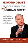
Howard Dean's Prescription for Real Healthcare Reform
by Howard Dean Igor Volsky Faiz ShakirAmericans have pondered how to reform healthcare since the days of Harry Truman. But for most Americans, little has changed-except that healthcare costs have soared, health insurance companies have grown bigger and more oppressive to both doctors and patients, and today even those Americans who pay dearly for health insurance frequently find that their policies don't adequately cover them when they need their coverage most. In his bold new book, Howard Dean-the physician and former governor widely credited for reviving the Democratic Party after the 2004 elections-tells Americans what needs to be done to successfully reform healthcare. One key, he writes, is to offer Americans the option to participate in a public health insurance program, much like Medicare. In this straight-talking guide to overcoming today's healthcare crisis, Dean spells out: * What Obama's healthcare plan is all about * How other countries handle healthcare * Which special interests are standing in the way of progress and why * How healthcare reform will help American businesses prosper * Why Americans need choice-between private and public health insurance coverage Millions of Americans lack health insurance; millions more pay for coverage that doesn't protect them from serious illness; and the status quo leaves Americans at the mercy of corporate interests. This persuasive argument from a passionate political strategist shows Americans how to take back the healthcare reins.

Howard Dean's Prescription for Real Healthcare Reform: How We Can Achieve Affordable Medical Care for Every American and Make Our Jobs Safer
by Howard DeanHoward Dean--the physician and former governor widely credited with reviving the Democratic Party after the 2004 elections--brings his unique, no-nonsense perspective to the healthcare debate. At a time when healthcare reform finally is within reach, Howard Dean's Prescription for Real Healthcare Reform is a riveting call to action that explains both the obstacles to reform and what it will take to achieve real healthcare reform.
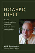
Howard Hiatt: How This Extraordinary Mentor Transformed Health with Science and Compassion (The\mit Press Ser.)
by Mark RosenbergThe seven-decade career of Howard Hiatt, a pioneer in public health, advocate for global health and health equity, a mentor to generations of healthcare leaders.Howard Hiatt—physician, scientist, advocate for global health, and mentor to generations of healthcare leaders—has spent much of his seven-decade career being ahead of his time. His innovative ideas as head of Harvard's School of Public Health from 1972 to 1984—about preventive medicine, the incorporation of cutting-edge science into the curriculum, and cross-disciplinary collaboration—met fierce resistance at the time but are now widely recognized building blocks of public health. Hiatt's interest in global health and health equity equipped him to advocate for a series of younger physicians and researchers, including Paul Farmer and Jim Kim, two founders of Partners in Health, and the prominent health policy expert Don Berwick. This book tells the story of Hiatt's life and work, with important lessons for today drawn from Hiatt's 92 years of experience.Hiatt, born in 1925, attended Harvard College and received an M.D. degree from Harvard Medical School. Before he headed the School of Public Health, he was a modernizing force as chief of medicine at Boston's Beth Israel Hospital. After his stormy tenure at SPH, he went to Brigham and Women's as a professor of medicine and a senior physician with a portfolio of his own devising. It was at the Brigham that Hiatt took on the role of mentor, influencing generations of physicians and staking out new territory in the fields of global health and clinical effectiveness. He is still active at 92 as teacher and mentor.
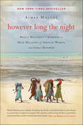
However Long the Night: Molly Melching's Journey to Help Millions of African Women and Girls Triumph
by Aimee MolloyIn However Long the Night, Aimee Molloy tells the unlikely and inspiring story of Molly Melching, an American woman whose experience as an exchange student in Senegal led her to found Tostan and dedicate almost four decades of her life to the girls and women of Africa.This moving biography details Melching's beginnings at the University of Dakar and follows her journey of 40 years in Africa, where she became a social entrepreneur and one of humanity's strongest voices for the rights of girls and women.Inspirational and beautifully written, However Long the Night: Molly Melching's Journey to Help Millions of African Women and Girls Triumph is a passionate entreaty for all global citizens. This book is published in partnership with the Skoll Foundation, dedicated to accelerating innovations from organizations like Tostan that address the world's most pressing problems.
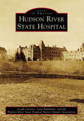
Hudson River State Hospital (Images of America)
by Joseph Galante Lynn Rightmyer the Hudson River State Hospital Nurses Alumni AssociationFor 141 years, Hudson River State Hospital was home to tens of thousands of individuals suffering from mental illness. The facility grew from a 208-acre parcel in 1871 with seven patients to 752 acres with five dozen separate buildings containing nearly 6,000 patients in 1954. The main building was constructed on a Kirkbride plan, a treating philosophy centered around an ornate building of equal proportions staffed by employees who integrated dignity and compassion into health care. Famous architects Frederick Clark Withers and Calvert Vaux drafted the main building in 1869. The landscape was penned by Frederick Law Olmstead, perhaps best known for the design of New York City's Central Park.
Hughes Syndrome
by Graham HughesThis second edition is a result from a worldwide surge of interest in antiphospholipid syndrome. This disease, also known as Hughes Syndrome, affects all organs of the body including the placenta, and therefore touches all areas of medicine. Many publications have shown that APS causes miscarriage, strokes, heart attacks and teenage epilepsy. This new edition will see many of the illustrations updated as well as an increase in the number of figures within the chapters, to help the reader understand and treat this disease. It aims to help patients with Hughes Syndrome and their families, as well as trainee doctors , GPs, nurses and midwives.
Hughes Syndrome: Highways and Byways
by Graham Hughes Munther A KhamashtaHughes Syndrome: Highways and Byways is a handy and easy-to-read guide to the main features of Hughes syndrome. There has been worldwide interest in this 'new' syndrome (first described in 1983). A clotting tendency which can potentially affect any organ in the body, it is: * responsible for 1 in 5 recurrent miscarriages (now successfully treated with aspirin and heparin) * responsible for 1 in 5 young (under 45) strokes * a major cause of early heart attacks * responsible for 1 in 5 deep vein thromboses * an important cause of migraine. Hughes Syndrome: Highways and Byways is a valuable resource for professionals with an interest in the subject, as well as general practitioners and patients.
Hughes Syndrome: The Antiphospholipid Syndrome
by Graham Hughes Shirish SangleHughes Syndrome: The Antiphospholipid Syndrome, A Guide for Students provides an in-depth analysis into the main effects of Hughes Syndrome. In 1983, Dr Graham Hughes, and his team in London, described a syndrome and subsequently developed simple blood tests to diagnose the condition. This syndrome is characterised by thrombosis (both in limbs and internal organs), headaches, memory loss, strokes and, in pregnant women, placental clotting and recurrent miscarriage. The syndrome, now known worldwide as Hughes Syndrome, or the Antiphospholipid Syndrome, is common - being responsible for example, for up to 1 in 5 cases of young stroke and more importantly, it is treatable. Hughes Syndrome: The Antiphospholipid Syndrome, A Guide for Students details the effects of Hughes Syndrome on the major organs, making it a valuable reference tool for students in training.
Hugo and Russell's Pharmaceutical Microbiology
by Brendan F. Gilmore Stephen P. DenyerHugo & Russell’s Pharmaceutical Microbiology Discover the very latest developments in pharmaceutical microbiology in the 9th edition of this popular textbook Microbiology is one of the essential pharmaceutical sciences upon which the study and practice of pharmacy is built. It has a bearing on all aspects of the manufacture of medicines and sterile products, from their design and development to their delivery as quality products. Few interventions are more central to modern medicine than the treatment of infection, where antibiosis, vaccination and hygienic practices have essential roles to play. The COVID-19 pandemic, the appearance of new pathogens and the rise of antibiotic resistance have demonstrated most completely the need for pharmaceutical practitioners, researchers and industrial scientists to be fully conversant with this field. The 9th edition of Hugo and Russell’s Pharmaceutical Microbiology has been updated to meet this need. Having long served as the sole comprehensive textbook covering this subject, it has now been adapted to a critical new period in the advancement of medical and pharmaceutical research and development. Its experienced editors have incorporated contributions from subject experts and created a text which will serve the next generation of pharmacy students, pharmaceutical industry scientists and researchers. In this ninth edition of Hugo and Russell’s Pharmaceutical Microbiology, readers will find: A mix of established and new authors bringing practical and research experience to their chapters Material covering the fundamentals of microbiology, microbial behavior and laboratory investigation Revised chapters incorporating new material on microbe-host interactions, antibiotic resistance, emerging pathogens, public health microbiology, healthcare-associated infection and pharmaceutical manufacture Emerging understandings from the COVID-19 pandemic on infection prevention and control and vaccine development Practitioners providing their insights on clinical practice and pharmaceutical production An accompanying website incorporating teaching resources Hugo and Russell’s Pharmaceutical Microbiology, 9th edition promises to remain the essential text for pharmacy and medical students, as well as researchers and industry professionals.
Human Abdominal Hydatidosis
by Ajaz Ahmad Malik Shams Ul BariThe book explores various aspects of hydatid disease, including the background, parasitology, epidemiology, etiology, pathogenesis and presentation in humans. It features dedicated chapters on hydatidosis of liver, spleen, peritoneum, kidney, pelvis, and disseminated hydatid disease, and also provides detailed information on the latest surgical and non-surgical methods for treating the condition, such as drug therapy and laproscopic management. The book is primarily intended for undergraduate and postgraduate students of surgery and medicine, but is also useful to veterinary science students and pharmaceutical companies. Further, it serves as a valuable reference resource for academics and researchers in associated fields.
Human Activity Recognition Challenge (Smart Innovation, Systems and Technologies #199)
by Sozo Inoue Md Atiqur Rahman Ahad Paula LagoThe book introduces some challenging methods and solutions to solve the human activity recognition challenge. This book highlights the challenge that will lead the researchers in academia and industry to move further related to human activity recognition and behavior analysis, concentrating on cooking challenge. Current activity recognition systems focus on recognizing either the complex label (macro-activity) or the small steps (micro-activities) but their combined recognition is critical for analysis like the challenge proposed in this book. It has 10 chapters from 13 institutes and 8 countries (Japan, USA, Switzerland, France, Slovenia, China, Bangladesh, and Columbia).
Human Agency and Neural Causes: Philosophy of Action and the Neuroscience of Voluntary Agency
by J. D. RunyanHuman Agency and Neural Causes provides an analysis of our everyday thought about our conduct, and the neuroscience research concerning voluntary agency. J.D. Runyan argues that our findings through neuroscience are consistent with what would be expected if we are, in fact, voluntary agents.
Human Airway Inflammation
by Louise E. Donnelly Duncan F. RogersThis book offers a wide range of readily reproducible methods for sampling, isolating, and culturing all of the major inflammatory cells involved in the pathophysiology of asthma and COPD. The collection of techniques detailed here involve biopsy and sampling of airway liquids such as sputum and exhaled gases such as nitric oxide. Also provided is a full range of methods for the isolation and characterization of cells and for the measurement of the major inflammatory markers, along with protocols for measuring many of the inflammatory mediators and enzymes released during lung inflammation. Among the more advanced techniques are the measurement of exhaled hydrocarbons and F2 isoprostanes, granulocyte pharmacodynamics in whole blood measured by flow cytometry, and tracing mediator trafficking in eosinophils using confocal microscopy. Comprehensive and highly practical, the methods presented in Human Airway Inflammation: Sampling Techniques and Analytical Protocols provide today's basic and clinical researchers all the major techniques for investigating airway inflammation, and powerfully illuminate many novel targets for emerging drugs.
Human Anatomy
by Valerie O'Loughlin Michael McKinley Ronald Harris Elizabeth Pennefather-O'BrienWith its unrivaled art program and accessible writing style, McKinley et al.'s Human Anatomy stands apart from other anatomy texts. High-quality photographs paired with brilliantly rendered illustrations help students visualize, understand, and appreciate the wonders of human anatomy. <p><p> The author team incorporates their over seventy years of teaching experience into student-friendly Learning Strategies, Clinical View boxes, and progressive question sets that motivate students to internalize and apply what they've learned. Users who purchase Connect Plus receive access to the full online eBook version of the textbook, as well full access to LearnSmart, SmartBook, and Anatomy & Physiology Ӏ REVEALED.
Human Anatomy
by Valerie O'Loughlin Michael McKinley Elizabeth Pennefather-O'BrienHuman Anatomy stands apart from other texts as it guides students on a clearly written and expertly illustrated beginner’s path through the human body. High-quality photographs paired with brilliantly rendered illustrations help students visualize, understand, and appreciate the wonders of human anatomy. The author team incorporates their combined 70 years of teaching experience into student-friendly learning strategies, built around a pedagogical framework designed to foster retention and encourage the application of knowledge and understanding.
Human Anatomy
by Frederic H. Martini Michael J. Timmons Robert B. TallitschThe Eighth Edition includes new one- and two-page Spotlight Figures that seamlessly integrate text and visuals to guide students through complex topics. This program presents a better teaching and learning experience.
Human Anatomy
by Inc. Frederic H. Martini Robert B. Tallitsch Judi NathFor courses in one-semester human anatomy. <p><p> A fresh voice strengthens a visually compelling, classic human anatomy text. <p> Revered for its groundbreaking, atlas-style format, detailed illustrations, and exceptionally clear photographs, Human Anatomy has long been known for its quality and sophistication, as well as its scope and level of coverage. Accomplished author and award-winning educator Judi Nath joins the writing team, bringing a fresh voice, enhanced accuracy, and a clear, engaging writing style to the 9th Edition. <p><p> The new edition builds upon the legacy of previous editions and includes expanded clinical content to help students focus on the types of diseases and injuries they are likely to encounter in their future careers; visually stunning Spotlight Figures in every chapter to guide students through complex topics; and new features that focus students on the key point in each section and use simple analogies and memory devices to help students remember facts and concepts.