- Table View
- List View
Imaging and Diagnosis in Pediatric Brain Tumor Studies
by Monika Warmuth-MetzThis book describes the features in structural imaging of the most important pediatric brain tumors with the aim of enabling radiologists to make the correct differential diagnosis and to provide the pediatric oncologist with all the imaging information relevant to further management. The opening chapter is devoted to the complex subject of pediatric trials at the national and international levels and the importance of staging for stratification, differential treatment, and outcome. A general imaging protocol for children with brain tumors is presented, and individual chapters then identify key points for the differential diagnosis and staging of posterior fossa tumors, low- and high-grade gliomas, germ cell tumors, and craniopharyngiomas. The relevance of aspects such as tumor site and age to diagnosis is explained, and pitfalls associated with meningeal dissemination and treatment-related complications mimicking recurrence are highlighted. The importance of ensuring comparability of follow-up by use of standard MR (or CT) imaging is emphasized. In drawing on the lessons gained both from pediatric trials and from the author's own experience, this book will be invaluable for all radiologists.
Imaging and Focal Therapy of Early Prostate Cancer
by Mahdi MottaghiThis book encompasses an up-to-date, comprehensive review of the state-of-the-art for prostate gland preserving therapies. It provides insight into the latest research and clinical applications of image-guided diagnosis and minimally invasive focal, gland-preserving treatment for prostate cancer. Fully updated and revised, this text evaluates the scientific evidence for the evolving trend to treat intermediate risk, clinically localized prostate cancer in a focally ablative manner with novel gland-preserving, image-targeted therapy methods. Imaging and Focal Therapy of Early Prostate Cancer, Third Edition opens with a discussion of why patients and clinicians should consider focal therapy, then moves on to consider the question of active surveillance versus focal therapy from a global perspective, with chapters on North American, European, Southeast Asian, and South American perspectives. From there, chapters cover the scientific foundation of focal therapy, current and new approaches to image cancer foci within the prostate (multiparametric ultrasonography, multiparametric magnetic resonance imaging, etc.) and various biopsy techniques. Following this is detailed coverage of patient selection, treatment strategy, adjuvants to enhance therapy, outcomes, and patient centered interests, followed by a discussion of the strengths and limitations of various therapeutic modalities, such as cryotherapy, high intensity focused ultrasound, and photodynamic therapy, follows. The final sections of the book cover the assessment of focal therapy outcomes and look forward to the future of focal therapy for prostate cancer. Written by experts in the field and lavishly illustrated with detailed line-art and photographs, this text is designed as a comprehensive resource for urologists, radiation oncologists, medical oncologists, radiologists, uropathologists, molecular biologists, biomedical engineers, residents, fellows, nurses and allied professionals, and researchers with an interest in the diagnosis and novel targeted treatment of prostate cancer.
Imaging and Focal Therapy of Early Prostate Cancer
by Thomas J. PolascikImaging and Focal Therapy of Early Prostate Cancer evaluates the scientific evidence for the evolving trend to treat low to intermediate risk, clinically localized prostate cancer in a focally ablative manner with novel gland-preserving, focal therapy methods. Various ablative devices such as high intensity focused ultrasound, irreversible electroporation, photodynamic therapy, cryotherapy and laser ablation, among others, are discussed in regard to their strengths and limitations as a therapeutic modality. Emphasis is placed on tumor stage shift towards early stage disease with an increase in unilateral versus bilateral cancers validated by final pathology assessment of large prostatectomy series. Current and new approaches to image cancer foci within the prostate (3-Dimensional contrast-enhanced transrectal ultrasonography, multiparametric magnetic resonance image with spectroscopy, ETC) are presented along with biopsy techniques to map prostate cancer. Patient selection, treatment strategy, outcomes and safety concerns that may provide acceptable cancer control and improved quality of life for patients are all covered in detail. Written by experts in the field and lavishly illustrated with detailed line-art and photographs, Imaging and Focal Therapy of Early Prostate Cancer is a resourceful volume beneficial to practitioners specializing in the treatment and management of prostate cancer.
Imaging and Focal Therapy of Early Prostate Cancer
by Thomas J. PolascikThis text encompass an up-to-date, comprehensive review of the state-of-the-art for gland preserving therapies. Fully updated and revised, this text evaluates the scientific evidence for the evolving trend to treat intermediate risk, clinically localized prostate cancer in a focally ablative manner with novel gland-preserving, focal therapy methods. Various ablative devices such as high intensity focused ultrasound, irreversible electroporation, photodynamic therapy, cryotherapy and laser ablation, among others, is discussed in regard to their strengths and limitations as a therapeutic modality. Emphasis is placed on patient selection and outcomes utilizing both advanced imaging techniques and pathologic evaluation. Current and new approaches to image cancer foci within the prostate (multiparametric ultrasonography, multiparametric magnetic resonance image, etc) are presented along with various biopsy techniques, including robotics to map prostate cancer. Patient selection based on imaging and genomic classification, adjuvants to enhance therapy, treatment strategy, outcomes and patient centered concerns is discussed, providing an acceptable balance between cancer control and improved quality of life for patients. Written by experts in the field and lavishly illustrated with detailed line-art and photographs, Imaging and Focal Therapy of Early Prostate Cancer, Second Edition is designed as a comprehensive resource for urologists, radiation oncologists, medical oncologists, radiologists, uropathologists, molecular biologists, biomedical engineers, other clinicians -- residents, fellows, nurses and allied professionals -- and researchers with an interest in the diagnosis and novel treatment of prostate cancer. It will provide insight into the latest research and clinical applications of image-guided diagnosis and minimally invasive focal, gland-preserving treatment for prostate cancer.
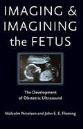
Imaging and Imagining the Fetus: The Development of Obstetric Ultrasound
by Malcolm Nicolson John E. FlemingHow engineers and clinicians developed the ultrasound diagnostic scanner and how its use in obstetrics became controversial.To its proponents, the ultrasound scanner is a safe, reliable, and indispensable aid to diagnosis. Its detractors, on the other hand, argue that its development and use are driven by the technological enthusiasms of doctors and engineers (and the commercial interests of manufacturers) and not by concern to improve the clinical care of women. In some U.S. states, an ultrasound scan is now required by legislation before a woman can obtain an abortion, adding a new dimension to an already controversial practice. Imaging and Imagining the Fetus engages both the development of a modern medical technology and the concerted critique of that technology.Malcolm Nicolson and John Fleming relate the technical and social history of ultrasound imaging—from early experiments in Glasgow in 1956 through wide deployment in the British hospital system by 1975 to its ubiquitous use in maternity clinics throughout the developed world by the end of the twentieth century. Obstetrician Ian Donald and engineer Tom Brown created ultrasound technology in Glasgow, where their prototypes were based on the industrial flaw detector, an instrument readily available to them in the shipbuilding city. As a physician, Donald supported the use of ultrasound for clinical purposes, and as a devout High Anglican he imbued the images with moral significance. He opposed abortion—decisions about which were increasingly guided by the ultrasound technology he pioneered—and he occasionally used ultrasound images to convince pregnant women not to abort the fetuses they could now see.Imaging and Imagining the Fetus explores why earlier innovators failed where Donald and Brown succeeded. It also shows how ultrasound developed into a "black box" technology whose users can fully appreciate the images they produce but do not, and have no need to, understand the technology, any more than do users of computers. These "images of the fetus may be produced by machines," the authors write, "but they live vividly in the human imagination."
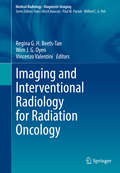
Imaging and Interventional Radiology for Radiation Oncology (Medical Radiology)
by Vincenzo Valentini Regina G. H. Beets-Tan Wim J. G. OyenThis book, edited by leading experts in radiology, nuclear medicine, and radiation oncology, offers a wide-ranging, state of the art overview of the specifics and the benefits of a multidisciplinary approach to the use of imaging in image-guided radiation treatments for different tumor types. The entire spectrum of the most important cancers treated by radiation are covered, including CNS, head and neck, lung, breast, gastrointestinal, genitourinary, and gynecological tumors. The opening sections of the book address background issues and a range of important technical aspects. Detailed information is then provided on the use of different imaging techniques for T staging and target volume delineation, response assessment, and follow-up in various parts of the body. The focus of the book ensures that it will be of interest for a multidisciplinary forum of readers comprising radiation oncologists, nuclear medicine physicians, radiologists and other medical professionals.
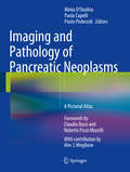
Imaging and Pathology of Pancreatic Neoplasms
by Paolo Pederzoli Mirko D'Onofrio Paola CapelliAlthough interest in pancreatic pathology is very high in the radiological and gastroenterological communities, it is still the case that less is known about pathology of the pancreas than about liver pathology, for example. Diagnosis depends on the structure of the pancreatic lesion, which can be directly visualized on US, CT or MR images. This atlas, which encompasses both the imaging and the pathology of pancreatic neoplasms, will therefore be invaluable in enabling radiologists and sonographers to understand the underlying pathology and in allowing pancreatic pathologists to understand the imaging translation. The emphasis in the atlas is very much on the pathological and imaging appearances, with most of the text concentrated at the beginning of the chapters. A comprehensive overview is provided of typical and atypical presentations and diverse aspects of common and uncommon pancreatic neoplasms, including ductal adenocarcinoma, neuroendocrine neoplasms, intraductal papillary mucinous neoplasms, cystic neoplasms, metastases and lymphoma.
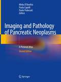
Imaging and Pathology of Pancreatic Neoplasms: A Pictorial Atlas
by Paolo Pederzoli Paola Capelli Mirko D’OnofrioThe second edition of this atlas focuses on imaging methods and techniques, new diagnostic concepts and therapeutic approaches in management of pancreatic neoplasms. Although interest in pancreatic pathology is very high in the radiological and gastroenterological communities, less is known about it than about, for example, liver pathology. Diagnosis depends on the structure of the pancreatic lesion, which can be directly visualized in US, CT or MR images.The book’s focus is very much on the imaging and pathological appearances, with most of the text concentrated at the beginning of the book followed by images gallery. A comprehensive overview is provided of typical and atypical presentations and diverse aspects of common and rare pancreatic tumors, including ductal adenocarcinomas with dedicated chapter to ductal adenocarcinoma downstaging, neuroendocrine neoplasms, cystic pancreatic neoplasms and intraductal papillary mucinous neoplasms. The Verona “Pancreas Centre,” the first institute of its kind in Italy and one of very few in the world, pursues an interdisciplinary approach to treating the problems of this organ, focusing on the patient, on research, and on teaching. A dedicated center that can look back on 40 years of tradition, many of its respected specialists have made essential contributions in surgery, gastroenterology, oncology, pathology, and radiology. Given its scope, this atlas will be an invaluable asset, helping radiologists understand the underlying pathology and helping pancreatic pathologists understand the imaging translation.
Imaging and Quantifying Neuronal Autophagy (Neuromethods #171)
by Esther Wong Ben LoosThe volume aims to explore the dynamic nature of the autophagy pathway, and the latest techniques that allow researchers to capture and quantify this process in neurons. The chapters in this volume cover topics such as fundamental, historical, and functional approaches that began in baker’s yeast; Saccharomyces cerevisiae; the role of both electron microscopy and live-cell imaging using fluorescently tagged autophagy proteins; and the rate of puncta appearance and its correlation with the rate of autophagosome formation. In the Neuromethods series style, chapters include the kind of detail and key advice from the specialists needed to get successful results in your laboratory. Cutting-edge and practical, Imaging and Quantifying Neuronal is a valuable resource that provides insights into the power of microscopy tools, live-cell imaging, and photoactivation and correlative techniques.
Imaging and Technology in Urology
by Steve Payne Sotonye Tolofari Dora Moon Benjamin StarmerThis book offers a new edition of the hugely successful title, Imaging & Technology in Urology--Principles and Clinical Applications edited by Steve Payne, Ian Eardley, Kieran O'Flynn in 2012. Essential reading for preparation of exit exams in Urology, it is used worldwide by exam candidates. Fully updated in essential areas of the book following on from recent developments in the last decade, it helps give preparation to candidates. The most comprehensive and reliable source of information on this particular topic.
Imaging and Technology in Urology
by Steve Payne Kieran O'Flynn Ian EardleyImaging and Technology: Principles and Clinical Applications is a practical and user-friendly consolidated source book for urologists, and urologists in training, regarding the basic science of imaging modalities used on a day-to-day basis in urological practice. Similarly, the intention is to provide an introduction to the technology that is used in the practice of urological surgery and the management of urological patients in the clinical setting. This knowledge level is appropriate for certification for independent consultant practice in urology in the UK. The book is also valuable to urologists and urological trainees outside of the UK and in other surgical specialities.
Imaging and Tracking Stem Cells: Methods and Protocols (Methods in Molecular Biology #2150)
by Kursad TurksenThis fully updated book brings together protocols to arm stem cell biologists with tools and approaches to continue uncovering the intricacies and regulatory mechanisms underlying stem cell biology. Through various models and organ systems, the volume reflects the numerous recent advances in cell lineage and lineage tracking. Written for the highly successful Methods in Molecular Biology series, chapters include introductions to their respective topics, lists of the necessary materials and reagents, step-by-step, readily reproducible laboratory protocols, and tips on troubleshooting and avoiding known pitfalls. Authoritative and up-to-date, Imaging and Tracking Stem Cells: Methods and Protocols, Second Edition is an ideal guide for novices and experts alike who are working to expand our knowledge in the field of stem cells.
Imaging and Urodynamics of the Lower Urinary Tract
by Uday PatelThis book is a practical text covering the imaging techniques and the range of urodynamic problems the trainee and practising urologist is likely to encounter including; congenital abnormalities of the bladder, functional abnormalities of the bladder, intraluminal abnormalities and staging of bladder cancer. The urethra and lower urinary tract are also dealt with. Although these disorders are covered in the major reference works, there is a need for a practical, accessible, user friendly text which provides clear and unambiguous guidelines on the best practice available.
Imaging and Visualization in The Modern Operating Room
by Yuman Fong Pier Cristoforo Giulianotti Jason Lewis Bas Groot Koerkamp Thomas ReinerThis text provides a state of the art overview of tools for guiding surgeons in the modern operating room. The text explains how many modalities in the current armamentarium of radiologic imaging have been brought to the operating room for real time use. It also explains the current use of near infrared, fluorescent, and chemo-luminescent imaging to guide minimally invasive and open surgery to improve outcome. The book is separated into two sections. The first, discusses the biologic principles that underlie novel visualization of normal organs and pathology. The currently available equipment and equipment anticipated in the near future is covered. The second section summarizes current clinical applications of advanced imaging and visualization in the OR. Novel means of visualizing normal anatomic structures such as nerves, bile duct, and vessels that enhance safety of many operations are covered. Novel biologic imaging using radio-labeled and fluorescent-labeled molecular probes that allow identification of inflammation, vascular abnormalities, and cancer are also discussed. Authored by scientists who pioneer research in optics and radiology, tool makers who use this knowledge to make surgical equipment, and surgeons who innovate the field of surgery using these new operative tools, Imaging and Visualization in the Modern Operating Room is a valuable guide for surgeons, residents and fellows entering the field.
Imaging for Patient-Customized Simulations and Systems for Point-of-Care Ultrasound: International Workshops, BIVPCS 2017 and POCUS 2017, Held in Conjunction with MICCAI 2017, Québec City, QC, Canada, September 14, 2017, Proceedings (Lecture Notes in Computer Science #10549)
by M. Jorge Cardoso, Tal Arbel, João Manuel R.S. Tavares, Stephen Aylward, Shuo Li, Emad Boctor, Gabor Fichtinger, Kevin Cleary, Bradley Freeman, Luv Kohli, Deborah Shipley Kane, Matt Oetgen and Sonja PujolThis book constitutes the refereed joint proceedings of the International Workshop on Bio-Imaging and Visualization for Patient-Customized Simulations, BIVPCS 2017, and the International Workshop on Point-of-Care Ultrasound, POCUS 2017, held in conjunction with the 20th International Conference on Medical Imaging and Computer-Assisted Intervention, MICCAI 2017, in Québec City, QC, Canada, in September 2017. The 12 full papers presented at BIVPCS 2017 and the 7 full papers presented at POCUS 2017 were carefully reviewed and selected. The papers feature research from complementary fields such as signal and image processing, mechanics, computational vision, mathematics, physics, informatics, computer graphics, bio-medical-practice, psychology and industry as well as ultrasound image systems applications.
Imaging for Pediatricians
by Antonio Martínez-Valverde Luisa Ceres-Ruiz María I. Martínez-LeónThe book, written by expert pediatricians and radiologists, is divided into ten chapters covering neurology, cardiology, neonatology, interventional radiology, the abdomen, the musculoskeletal system, the thorax, the genitourinary system, the fetus, and emergencies. Each chapter comprises ten cases that are presented in a standard way. After discussion of the disorder in question, four representative images are displayed and described with special attention to distinctive features. In addition, informative key references are provided, including a book or book chapter, a web link, and ten recent articles.
Imaging for Plastic Surgery
by Luca Saba Warren M. Rozen Alberto Alonso-Burgos Diego RibuffoPreoperative imaging is increasingly being adopted for preoperative planning in plastic and reconstructive surgery. Accurate preoperative analysis can reduce the length of operations and maximize surgical design and dissection techniques. Imaging for Plastic Surgery covers the techniques, applications, and potentialities of medical imaging technology in plastic and reconstructive surgery. Presenting state-of-the-art research on evolving imaging modalities, this cutting-edge text: Provides a practical introduction to imaging modalities that can be used during preoperative planning Addresses imaging principles of the face, head, neck, breast, trunk, and extremities Identifies the strengths and weaknesses of all available imaging modalities Demonstrates the added value of imaging in different clinical scenarios Comprised of contributions from world-class experts in the field, Imaging for Plastic Surgery is an essential imaging resource for surgeons, radiologists, and patient care professionals.
Imaging for Reconstructive Microsurgery
by Joon Pio Hong Giuseppe Visconti Akitatsu Hayashi Bernard T. LeeImaging in Reconstructive Microsurgery represents the first book in its kind the the Plastic Surgery and Reconstructive Microsurgery Community. This books includes all the imaging modalities available for plastic and reconstructive surgeons which adds major steps in the daily clinical practice. Most of these imaging modalities can and should be used by the operating surgeon her/himself to exponentially enhance and empower the clinical practice. This book provides step-by-step description of all the state-of-art imaging modalities for both perforator flap and lymphatic surgery, with the aim to provide a daily reference to colleagues who are novel to these procedures and for those who are looking forward to improve their practice. Encorporating imaging technology in clinical practice represents a paradigm shift in daily clinical practice with major enhancement of safety, minimal invasiveness and creativity finally leading to a next generation reconstructive approach. We believe that those imaging modalities will guide us to the future of microsurgery and super- microsurgery. This book will be the beginning of reader’s new journey.
Imaging for Students
by David Lisle Craig Hacking'I would have found this book invaluable at medical school, but as a now qualified GP I think it is a fabulous resource. The fact it covers so much is remarkable … It is so comprehensive – great images, well explained.' Donna Pilkington, GP with an interest in medical education, Northern Ireland, UK 'It is direct and succinct. Just what you need in a portable book that aims to give you the essentials [it does] a great job of incorporating a huge amount of information covering the wide range of radiology examinations and procedures into a readable and practical book for students. A good introduction for year 1 radiology residents too.' Dr Mike Hurrell, Clinical Senior Lecturer and Consultant Radiologist, University of Otago, Christchurch, New Zealand 'The text works very well for the Medical Imaging Students – providing an overview of each modality and key insights into the clinical question to be resolved … the information is presented in an accessible fashion and well-illustrated.' Associate Professor Debbie Starkey, Discipline Leader, Medical Radiation Sciences, School of Clinical Sciences, Faculty of Health, Queensland University of Technology, Brisbane, Australia Imaging for Students delivers step-by-step guidance to the range of imaging techniques available, providing a clear explanation of how each imaging modality actually works, and including information on the associated risks and hazards. Throughout, the importance of patient preparation and post-procedure observation is emphasized. Taking information from evidence-based studies and published guidelines, in line with current clinical practice, the book takes a highly logical approach to the investigation of clinical scenarios, where possible indicating the 'best first test'—vital to both appropriate clinical and cost-effective decision-making. Key Features: Readable and concise – focusing on the common diseases that medical students most frequently encounterFully revised and updated – including up-to-date information on the latest imaging techniques including spectral CT, liver elastography, new and emerging PET techniques, multiparametric imaging and the role of AIHeavily illustrated - over 450 high-quality photographs, many new to this edition including colour images, are essential to support this visual subjectHighly structured and accessible format – plentiful use of tables and lists, and introduction of new summary boxes, all ideal for study and exam preparationCompanion website – image library including normal anatomy, clinical cases and MCQs for self-assessment, RADS reporting systems and detailed staging systems for common tumours relevant to each section; visit www.routledge.com/cw/hacking Drawing on the extensive clinical and teaching experience of its respected author team, the fifth edition of Imaging for Students gives students and junior doctors everything they need to understand the advantages, disadvantages, and possible side effects of the imaging modalities available, and how to apply them appropriately in clinical practice. The Authors: Craig Hacking is an Associate Professor of Radiology and Academic Lead for Clinical Radiology, University of Queensland Medical School and a Consultant Radiologist and the Medical Director of Medical Imaging at the Royal Brisbane and Women’s Hospital, Brisbane, Australia. David Lisle is an Associate Professor of Medical Imaging, University of Queensland Medical School, a Consultant Radiologist at Brisbane Private Hospital, Brisbane, Australia, an examiner for the Royal Australian and New Zealand College of Radiologists and is the author of previous editions of Imaging for Students. </
Imaging for Students
by David A LisleImaging for Students delivers step-by-step guidance to the range of imaging techniques available, providing a clear explanation of how each imaging modality actually works, and including information on the associated risks and hazards. Throughout, the importance of patient preparation and post-procedure observation is emphasized.Taking information
Imaging from Cells to Animals In Vivo (Series in Cellular and Clinical Imaging)
by Margarida M. Barroso and Xavier IntesThis book offers an overview of imaging techniques used to investigate cells and tissue in their native environment. It covers the range of imaging approaches used, as well as the application of those techniques to the study of biological processes in cells and whole tissues within living organisms.
Imaging in CNS Drug Discovery and Development
by Edward Bullmore Richard J. Hargreaves David Borsook Lino R. BecceraDrug development today needs to balance agility, speed, and risk in defining probability of success for molecules, mechanisms, and therapeutic concepts. New techniques such as fMRI promise to be part of a sequence that could transform drug development. Although numerous review articles exist that discuss the use of imaging in drug development, no one source is available that combines the various techniques and includes a discussion of disease mapping. Imaging in CNS Drug Discovery and Development, Implications for Disease and Therapy will serve to distill the most salient developments in the use of imaging in drug development and disease mapping. It will launch evolving concepts that integrate new imaging technologies and paradigms with molecular medicine and molecular profiling ("monics") as well as consider the ethical issues that arise as a result of disease or state diagnosis and the use of imaging in the public eye.
Imaging in Cellular and Tissue Engineering
by Hanry Yu Nur Aida Abdul RahimDetails on specific imaging modalities for different cellular and tissue engineering applications are scattered throughout articles and chapters in the literature. Gathering this information into a single reference, Imaging in Cellular and Tissue Engineering presents both the fundamentals and state of the art in imaging methods, approaches, and app
Imaging in Clinical Neurosciences for Non-radiologists: An Atlas
by Hemanshu Prabhakar S. Leve DevarajanThis Atlas presents both normal and pathological conditions of the Brain and Spine pictorially. Targeted towards non-radiologists, it is a unique book with well labeled and self-explanatory images. All routine conditions involving neuroradiology have been included. Images from different radiological modalities such as X-ray, Computed Tomography (CT), Magnetic Resonance Imaging (MRI) and Digital Subtraction Angiography (DSA) have also been included. This book aims to serve as a ready reckoner for clinicians, trainees, residents as well as professional radiologists. Key Features Discusses topics related to allied branches of neurology, neuroanesthesia, neurointensive care and neurosurgery Presents both common and uncommon neurological conditions Contains actual real-life scans and images Works as a unique, quick reference guide of neuroradiological images for non-radiologists
Imaging in Clinical Oncology
by Athanasios D. Gouliamos John Andreou Paris KosmidisThis encompassing book is designed to contribute to a teamwork approach by promoting understanding between radiologists and clinical oncologists. All of the currently available imaging modalities of relevance in clinical oncology are covered, and the presentation of a broad spectrum of oncologic diseases (of most organ systems) on these modalities is discussed and illustrated. The role of multiparametric and multimodality imaging approaches providing both morphologic and functional information is considered in detail, and careful attention is paid to the latest developments in higher field (3T) MR imaging and advanced MR techniques such as diffusion-weighted imaging, diffusion tensor imaging, perfusion-weighted imaging and spectroscopy. The major challenge of incorporating progress in quantitative imaging technology into radiotherapy treatment planning, guidance, and monitoring is also addressed. This book will assist in refining the treatment approach in various oncologic diseases and organ systems based on specific imaging features. It will be of value to radiologists, oncologists, and other medical professionals involved in the diagnosis and treatment of oncology patients.