- Table View
- List View
MRI from A to Z
by Gary LineyThis second edition of Gary Liney's MRI from A-Z, much expanded from the first edition, is both a reflection of and an apt companion for the dramatic growth of the field of MRI. The MRI-trainee to the most seasoned practitioner in MRI will find this A-Z of the field, with 1,300 entries and 100 illustrations, an indispensable reference tool. Providing the reader with concise, clear and eloquent definitions of MRI terminology, this book is both highly practical and a pleasure to read.
MRI from Picture to Proton
by Moore Martin J. Elizabeth A. Donald W. Mcrobbie GravesMRI from Picture to Proton presents the basics of MR practice and theory in a unique way: backwards! The subject is approached just as a new MR practitioner would encounter MRI: starting from the images, equipment and scanning protocols, rather than pages of physics theory. The reader is brought face-to-face with issues pertinent to practice immediately, filling in the theoretical background as their experience of scanning grows. Key ideas are introduced in an intuitive manner which is faithful to the underlying physics but avoids the need for difficult or distracting mathematics. Additional explanations for the more technically inquisitive are given in optional secondary text boxes. The new edition is fully up-dated to reflect the most recent advances, and includes a new chapter on parallel imaging. Informal in style and informed in content, written by recognized effective communicators of MR, this is an essential text for the student of MR.
MRI in Epilepsy
by Horst UrbachMRI can play an important role in identifying and localizing epileptogenic foci. This book aims to provide the clinical and imaging information required in order to decide whether an MRI scan is appropriate and whether it is likely to be sufficient to detect a lesion. The first part of the book presents background information on epilepsy patients and explains how to perform an MRI examination. Detailed attention is paid to functional MRI and post-processing, and the examination of subcategories of patients is also discussed. The second part of the book then documents the MRI findings obtained in the full range of epileptogenic lesions with the aid of high-quality images. Throughout, emphasis is placed on guiding the reader in the correct interpretation of the imaging findings. Both radiologists and referring physicians will find this book to be an indispensable guide to the optimal use of MRI in epilepsy.
MRI in Practice
by Carolyn Kaut Roth John Talbot Catherine WestbrookSince the first edition was published in 1993, MRI in Practice has become the standard text for radiographers, technologists, radiology residents, radiologists and even sales representatives on the subject of Magnetic Resonance Imaging (MRI). This text is essential reading on undergraduate and postgraduate MRI courses. Furthermore MRI in Practice has come to be known as the number one reference book and study guide in the areas of MR instrumentation, principles, pulse sequences, image acquisition, and imaging parameters for the advanced level examination for MRI offered by the American Registry for Radiologic Technologists (ARRT) in the USA.The book explains in clear terms the theory that underpins magnetic resonance so that the capabilities and operation of MRI systems can be fully appreciated and maximised. This fourth edition captures recent advances, and coverage includes: parallel imaging techniques and new sequences such as balanced gradient echo.Building on the success of the first three editions, the fourth edition has been fully revised and updated. The book now comes with a companion website at www.wiley.com/go/mriinpractice which hosts animated versions of a selection of illustrations in the book that are used on the MRI in Practice Course. These animations and accompanying text are aimed at helping the reader's comprehension of some of the more difficult concepts. The website also hosts over 200 interactive self-assessment exercises to help the reader test their understanding.MRI in Practice features:Full color illustrationsLogical presentation of the theory and applications of MRIA new page designA companion website at www.wiley.com/go/mriinpractice featuring interactive multiple choice questions, short answer questions PLUS animations of more complex concepts from the bookFor more information on the MRI in Practice Course and other learning resources by Westbrook and Talbot, please visit www.mrieducation.com
MRI in Practice
by John Talbot Catherine WestbrookMRI in Practice continues to be the number one reference book and study guide for the registry review examination for MRI offered by the American Registry for Radiologic Technologists (ARRT). This latest edition offers in-depth chapters covering all core areas, including: basic principles, image weighting and contrast, spin and gradient echo pulse sequences, spatial encoding, k-space, protocol optimization, artefacts, instrumentation, and MRI safety. The leading MRI reference book and study guide. Now with a greater focus on the physics behind MRI. Offers, for the first time, equations and their explanations and scan tips. Brand new chapters on MRI equipment, vascular imaging and safety. Presented in full color, with additional illustrations and high-quality MRI images to aid understanding. Includes refined, updated and expanded content throughout, along with more learning tips and practical applications. Features a new glossary. MRI in Practice is an important text for radiographers, technologists, radiology residents, radiologists, and other students and professionals working within imaging, including medical physicists and nurses.
MRI of Cardiovascular Malformations
by Bruno KastlerMRI is a non-invasive and non-ionizing imaging modality that is perfectly suited for the diagnosis and follow-up of both pediatric and adult congenital heart disease. It provides a large field of view and has the unique ability to depict complex cardiac and vascular anatomy and to measure cardiac function and flow within one examination. MRI is the ideal complement to echocardiography whenever the information provided by the latter is limited. This book has been conceived as a self-teaching manual that will assist qualified radiologists, cardiologists, and pediatricians, as well as those in training. It is richly illustrated with numerous images and drawings that cover all usual and most unusual anomalies. The principal author, Professor Bruno Kastler, is head of radiology at Besançon University Hospital, France and is board certified in both radiology and cardiology.
MRI of Degenerative Disease of the Spine
by Paola D'AprileThis richly illustrated case-based atlas thoroughly depicts the role of MR imaging in the assessment of patients presenting with pain due to degenerative disease of the spine and will serve as an excellent guide to differential diagnosis. Importantly, generic radicular compression is the main reason for the painful symptomatology in only a limited number of cases, and this book illustrates and emphasizes how various anatomic elements of the spine can be responsible. The imaging features of a range of disorders involving both the anterior and posterior elements of the spine are described, including active inflammatory osteochondrosis, atypical herniated discs, facet joint disorders, spondylolysis, and degenerative-inflammatory changes of the spinal ligaments and posterior perispinal muscles. Each example is supported by clinical data, and a series of unusual cases are also presented. MR study protocols include T2-weighted sequences with fat saturation and contrast-enhanced T1-weighted sequences with fat saturation to allow better visualization or highlighting of various inflammatory changes in the spine. Radiologists, neuroradiologists, neurosurgeons, orthopedists, and rehabilitation physicians will all find this atlas a valuable asset in their practice.
MRI of Degenerative Disease of the Spine: A Case-Based Atlas
by Paola D'Aprile Alfredo TarantinoThis is the second edition of an acclaimed, richly illustrated and comprehensive case-based atlas focusing on MRI of degenerative changes in the osteoarticular structures of the spine. Spinal degenerative disease is highly prevalent in the general population and its incidence increases with age. At the same time, degenerative spinal conditions are one of the most common causes of pain.The book presents a comprehensive overview of the MR findings observed in degenerative disease of spinal joints, ligaments and paravertebral muscles, and offers guidance on selecting the appropriate imaging protocol, which is critical in detecting the potentially very subtle changes. The MR study protocols presented include T2-weighted sequences with fat saturation and contrast-enhanced T1-weighted sequences with fat saturation, since these sequences permit better visualization of inflammatory changes of both anterior and posterior elements of the spine.This richly illustrated second edition highlights the inflammatory component of the degenerative pathology of the spine, which in most cases is responsible for the painful symptomatology. It also discusses in detail the use of contrast medium in MRI of spinal degenerative disease. The “case-based” structure of the atlas allows easy but effective consultation by radiologists, neuroradiologists, rheumatologists, orthopedists and physiatrists, as well as students.
MRI of Gynaecological Diseases: Illustrations and Cases
by Guofu ZhangThis book systematically covers MRI imaging findings of a spectrum of gynaecological diseases, including ovarian cysts, uterine fibroids, cervical cancer, and endometrial cancer, etc. All chapters are arranged in the same format: simply clinical history is firstly presented in order to help readers mimic the real clinical scenario, and then untypical features in many common diseases and typical features in many uncommon diseases are interpreted. In the end of each chapter, tips in differential diagnosis were summarized by analysing a class of cases with similar imaging performance. All cases in the book have the detailed pathological results, and the pathological specimens are also shown in some cases. It will be an ideal reference for physicians involved in the diagnosis and treatment of gynaecological diseases.
MRI of Rectal Cancer
by Mathias Langer Arnd-Oliver SchäferToday magnetic resonance imaging (MRI) of rectal cancer is one of the most interesting topics in radiology. Over the last decade technical refinements have strengthened the role of MRI as a first-line diagnostic strategy for accurate staging of rectal cancer. MRI is perfectly suited to predict tumor-free circumferential resection margins. Additionally, high accuracies have been reported for the T and N stages, which is comparable to transrectal ultrasound (TRUS). Novel approaches to moving table MRI serve as a platform for thoracic, abdominal and pelvic staging of rectal cancer patients within one examination. The broad spectrum of high-quality illustrations and the contribution of clinical experts provide substantial insight into the sophisticated field of rectal cancer management.
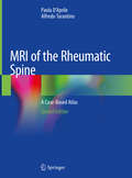
MRI of Rheumatic Spine
by Paola D'AprileThis richly illustrated and comprehensive case-based atlas documents the MR findings observed in spondyloarthritis and offers guidance on selection of the appropriate imaging protocol, which is critical in detecting the potentially very subtle changes. The presented MR study protocols include T2-weighted sequences with fat saturation and contrast-enhanced T1-weighted sequences with fat saturation, these being sequences which permit better visualization of inflammatory changes of both anterior and posterior elements of the spine. Cases of spondylitis, discitis, osteoarthritis and sacroiliitis are described and concise information is provided on the clinical history of the rheumatic diseases. The inclusion of a large number of high-resolution images ensures that the atlas will serve as a guide to differentiation between potentially confounding diseases and an aid to early diagnosis, which has become essential with the advent of new treatments in the field of spondyloarthritis (TNF inhibitors). In addition to radiologists, neuroradiologists, rheumatologists, orthopedists and physiatrists will greatly benefit from the contents of this volume and its thorough presentation of the rheumatic diseases.
MRI of Short and Ultrashort-T_2 Tissues: Making the Invisible Visible
by Graeme M. Bydder Jiang DuThis book comprehensively covers ultrashort echo time (UTE), zero echo time (ZTE), and other magnetic resonance imaging (MRI) acquisition techniques for imaging of short and ultrashort-T2 tissues. MRI uses a large magnet and radio waves to generate images of tissues in the body. The MRI signal is characterized by two time constants, spin-lattice relaxation time (T1) which describes how fast the longitudinal magnetization recovers to its initial value after tipping to the transverse plane, and spin-spin relaxation time (T2) which describes how fast the transverse magnetization decays. Conventional MRI techniques have been developed to image and quantify tissues with relatively long T2s. However, the body also contains many tissues and tissue components such as cortical bone, menisci, ligaments, tendons, the osteochondral junction, calcified tissues, lung parenchyma, iron containing tissues, and myelin, which have short or ultrashort-T2s. These tissues are “invisible” with conventional MRI, and their MR and tissue properties are not measurable. UTE and ZTE type sequences resolve these challenges and make these tissues visible and quantifiable.This book first introduces the basic physics of conventional MRI as well as UTE and ZTE type MRI, including radiofrequency excitation, data acquisition, and image reconstruction. A series of contrast mechanisms are then introduced and these provide high resolution, high contrast imaging of short and ultrashort-T2 tissues. A series of quantitative UTE imaging techniques are described for measurement of MR tissue properties (proton density, T1, T2, T2*, T1p,magnetization transfer, susceptibility, perfusion and diffusion). Finally, clinical applications in the musculoskeletal, neurological, pulmonary and cardiovascular systems are described.This is an ideal guide for physicists and radiologists interested in learning more about the use of UTE and ZTE type techniques for MRI of short and ultrashort-T2 tissues.
MRI of Tissues with Short T2s or T2*s
by Ian R. Young Gary D. Fullerton Graeme M. BydderThe content of this volume has been added to eMagRes (formerly Encyclopedia of Magnetic Resonance) - the ultimate online resource for NMR and MRI. Up to now MRI could not be used clinically for imaging fine structures of bones or muscles. Since the late 1990s however, the scene has changed dramatically. In particular, Graeme Bydder and his many collaborators have demonstrated the possibility - and importance - of imaging structures in the body that were previously regarded as being "MR Invisible". The images obtained with a variety of these newly developed methods exhibit complex contrast, resulting in a new quality of images for a wide range of new applications.This Handbook is designed to enable the radiology community to begin their assessment of how best to exploit these new capabilities. It is organised in four major sections - the first of which, after an Introduction, deals with the basic science underlying the rest of the contents of the Handbook. The second, larger, section describes the techniques which are used in recovering the short T2 and T2* data from which the images are reconstructed. The third and fourth sections present a range of applications of the methods described earlier. The third section deals with pre-clinical uses and studies, while the final section describes a range of clinical applications. It is this last section that will surely have the biggest impact on the development in the next few years as the huge promise of Short T2 and T2* Imaging will be exploited to the benefit of patients.In many instances, the authors of an article are the only research group who have published on the topic they describe. This demonstrates that this Handbook presents a range of methods and applications with a huge potential for future developments.About EMR Handbooks / eMagRes Handbooks The Encyclopedia of Magnetic Resonance (up to 2012) and eMagRes (from 2013 onward) publish a wide range of online articles on all aspects of magnetic resonance in physics, chemistry, biology and medicine. The existence of this large number of articles, written by experts in various fields, is enabling the publication of a series of EMR Handbooks / eMagRes Handbooks on specific areas of NMR and MRI. The chapters of each of these handbooks will comprise a carefully chosen selection of articles from eMagRes. In consultation with the eMagRes Editorial Board, the EMR Handbooks / eMagRes Handbooks are coherently planned in advance by specially-selected Editors, and new articles are written (together with updates of some already existing articles) to give appropriate complete coverage. The handbooks are intended to be of value and interest to research students, postdoctoral fellows and other researchers learning about the scientific area in question and undertaking relevant experiments, whether in academia or industry.Have the content of this Handbook and the complete content of eMagRes at your fingertips! Visit: www.wileyonlinelibrary.com/ref/eMagResView other eMagRes publications here
MRI of the Female and Male Pelvis
by Riccardo Manfredi Roberto Pozzi MucelliBased on the experience of two Italian referral centers, the book depicts the characteristic findings obtained when using MR imaging to study the male and female pelvis including the obstetric applications. Each chapter provides a comprehensive account of the use of the imaging technique of examination, including the most recent advances in MR imaging, the anatomy and MR possibilities in the identification, characterization and staging of the different pelvic diseases highlighting its diagnostic possibilities. The advances in fetal MRI, representing the cutting edge of pelvic MR imaging, will also be depicted. The text is complemented by numerous illustrations, as well as clinical cases that make this a very practice-oriented work, presenting the role of diagnostic imaging in every-day clinical activity. The volume will prove an invaluable guide for both residents and professionals with core interest in gynecology, obstetrics and urology.
MRI of the Gastrointestinal Tract
by Jaap StokerMRI has become an important tool in the management of patients with diseases of the gastrointestinal tract, such as rectal cancer and inflammatory bowel diseases. This book, written by distinguished experts in the field, discusses in detail the technical, practical, and clinical aspects of MRI of the gastrointestinal tract. The chapters on technique encompass the most recent developments and address such topics as contrast media, high field strength MRI, and perfusion MRI. Subsequently, individual chapters are devoted to the clinical applications of MRI in the different parts of the gastrointestinal tract. Both established applications and new frontiers are considered, with the aid of numerous high-quality illustrations. By combining chapters dedicated to technical aspects and clinically oriented chapters, this book will prove very instructive for the novice while simultaneously offering experienced practitioners further insights into the value of MRI of the gastrointestinal tract.
MRI of the Knee
by Nicolae V. Bolog Gustav Andreisek Erika J. UlbrichThis book is divided into chapters that cover MRI of all structures of the knee joint in the order that is usually used in practice - cruciate ligaments, collateral ligaments, menisci, cartilage, subchondral bone, patella, synovia, muscles and tendons, arteries, veins and bones With the aid of numerous images, each chapter provides comprehensive descriptions of the anatomy, the normal MR appearance, pathological MR findings, and postoperative MRI appearance. A text box at the end of each chapter clearly describes how the MRI report should be compiled and identifies what should be included when reporting on specific lesions. The book will be an ideal guide for radiologists and will also be relevant for orthopaedic surgeons, rheumatologists, and physiotherapists.
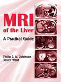
MRI of the Liver: A Practical Guide
by Philip J. Robinson Janice WardWith 2300 radiological images dispersed throughout the text, this source provides an expansive armamentarium of case studies and examples showcasing both common and uncommon liver pathologies. Serving as an unparalleled how-to source for the investigation of liver disease by MRI, this guide demonstrates key MRI techniques currently utilized in clin
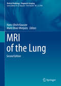
MRI of the Lung (Medical Radiology)
by Hans-Ulrich Kauczor Mark Oliver WielpützThis book provides a comprehensive overview of how to use MRI for the imaging of lung disease. Special emphasis is placed on routine applications and the clinical impact of MRI in each setting. In addition, current technological developments are reviewed and information presented on dedicated applications of MRI in preclinical and translational research, clinical trials, and specialized institutions.During the past two decades, significant advances in the technology have enabled MRI to enter and mature in the clinical arena of chest imaging. Standard protocols are now readily available on MR scanners, and MRI is recommended as the first- or second-line imaging modality for a variety of lung diseases, not limited to cystic fibrosis, pulmonary hypertension, and lung cancer. The benefits and added value of MRI originate from its ability to both visualize lung structure and provide information on different aspects of lung function, such as perfusion, respiratory motion, ventilation, and gas exchange. On this basis, novel quantitative surrogates for lung function and therapy control (imaging biomarkers) are generated. The second edition of MRI of the Lung has been fully updated to take account of recent advances. It is written by an internationally balanced team of renowned authors representing all major groups in the field.
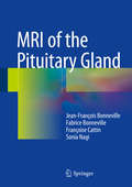
MRI of the Pituitary Gland
by Jean-François Bonneville Fabrice Bonneville Françoise Cattin Sonia NagiThis clinically oriented book will familiarize the reader with all aspects of the diagnosis of tumors and other disorders of the pituitary gland by means of magnetic resonance imaging (MRI). The coverage includes acromegaly, Cushing's disease, Rathke cleft cysts, prolactinomas, incidentalomas, nonsecreting adenomas, other lesions of the sellar area, hypophysitis, and central diabetes insipidus. Normal radiologic anatomy and the numerous normal variants are described, and guidance is also provided on difficulties, artifacts, and other pitfalls. The book combines concise text and high-quality images with a question and answer format geared toward the needs of the practitioner. MRI is today considered the cornerstone in the diagnosis of diseases of the hypophyseal-hypothalamic region but the relatively small size of the pituitary gland, its deep location, the many normal anatomic variants, and the often tiny size of lesions can hinder precise evaluation of the anatomic structures and particularly the pituitary gland itself. Radiologists and endocrinologists will find MRI of the Pituitary Gland to be full of helpful information on this essential examination, and the book will also be of interest to internists and neurosurgeons.
MRI of the Rheumatic Spine: A Case-Based Atlas
by Paola D'Aprile Alfredo TarantinoInflammatory pathology of the spine is an underestimated, but widespread condition among the population. This atlas shares essential information and case studies on key aspects of MRI for rheumatic inflammatory lesions of the osteoarticular structures of the spine, offering radiologists and clinicians a valuable guide to the management of patients with inflammatory back pain. In particular, this richly illustrated and comprehensive case-based atlas documents the MR findings observed in seronegative spondyloarthritis and rheumatoid arthritis of the cervical spine, providing guidelines on selecting the appropriate imaging protocol.The book is divided into two main parts, the first of which describes the general, clinical and radiological aspects of both spondyloarthritis and, as a new entry in the second edition, rheumatoid arthritis of the cervical spine. The second part then presents cases of spondylitis; discitis; facet joint and costovertebral osteoarthritis; sacroiliitis and rheumatoid arthritis of the atlanto-axial joint, including concise clinical and radiological information and a wealth of high-resolution images.The idea for a second edition of this book stemmed, on the one hand, from the global response to the first edition, and on the other, from a desire to expand the content to include wider illustrations and to address the clinically and socially relevant topic of the inflammatory pathology of the spine in more depth. The second edition offers a comprehensive guide to MRI of inflammatory diseases of the spine, facilitating an early diagnosis, which has become essential with the advent of new effective but expensive treatments with TNF inhibitors. Thanks to its “case-based” structure, the book offers an easy-to-use but thorough handbook for radiologists, neuroradiologists, rheumatologists, orthopedists and physiatrists, as well as students.
MRI of the Spine: A Guide for Orthopedic Surgeons
by William B. Morrison John A. Carrino Adam E. FlandersUtilizing plentiful radiological images to illustrate each topic, this text is a comprehensive and descriptive review of magnetic resonance imaging (MRI) interpretation for the spine, emphasizing standardized nomenclature and grading schemes. The book begins with current MR imaging protocols, including indication, sequencing and advanced imaging techniques, and a review of the relevant anatomy of the spine and its anomalies. Subsequent chapters encompass topics of trauma, degenerative disease, infection, inflammatory disease, as well as neoplastic and metabolic disease. Spinal cord and dural lesions will also be presented, with additional chapters dedicated to MRI evaluation of the post-operative patient. The format is reader-friendly, utilizing an efficient presentation of the essential principles and important findings on MR images of the spine, with a wealth of high-quality figures, graphics and tables for differential diagnosis as well as tips and tricks from experts in the field. Presenting the most up-to-date protocols and suggested interpretations, MRI of the Spine will be a solid reference for orthopedic surgeons, sports medicine specialists, neurosurgeons, radiologists and all clinicians and support staff caring for the spine.
MRI of the Temporomandibular Joint: Correlation Between Imaging and Pathology
by Tiziana Robba Carlotta Tanteri Giulia TanteriThis book is the outcome of a fruitful, long-standing cooperation between expert radiologists and clinicians, and explains the most relevant features and technical requirements that are needed to optimally conduct and assess MR examinations for temporomandibular joint (TMJ) pathologies. TMJ conditions are increasingly gaining attention, as the underlying diseases involved can vary considerably and be difficult to diagnose. Similarly, several imaging sub-specialties (e.g. dental radiology, neuroradiology, and musculoskeletal radiology) now find themselves dealing with the temporomandibular joints. The authors provide essential information on TMJ anatomy, dynamics, function and dysfunction. Correlations between clinical aspects and MRI findings are discussed and guidance for the correct interpretation of results is offered. Special findings that are helpful for differential diagnosis (arthritis, osteochondroma, synovial chondromatosis) are also examined. Given its extensive and varied coverage, the book offers a valuable asset for radiologists, dentists, gnathologists, maxillofacial surgeons, orthodontists and other professionals seeking a thorough overview of the subject
MRI of the Upper Extremity: Elbow, Wrist, and Hand
by Bethany U. CasagrandaThis book systematically discusses the anatomy and pathology of three specific regions of the upper extremity: the elbow, wrist, and hand. Divided into three sections, by body part, chapters cover anatomy and pathology. The anatomy chapters give a comprehensive view of each body part and normal variants found there. Although the primary modality emphasized will be MRI, illustrations and other modalities, including plain radiograph and CT, will be used to comprehensively discuss the anatomy of each region. Liberally illustrated, the pathology chapters then cover both traumatic and non-traumatic causes for imaging and detail how to perform and interpret each MRI. Specific examples include: osseous trauma, soft tissue trauma, and tumor imaging. Chapters are written with the deliberate intention to be of value to all levels of radiology training while remaining a reliable resource for attending radiologists.
MRI of the Whole Body: An Illustrated Guide for Common Pathologies
by Edward Hoey Nikhil Bhuskute Amit Lakkaraju Kshitij MankadThe optimal use of magnetic resonance imaging poses a constant challenge as the technology is continually and rapidly advancing. This leaves the MR practitioner, beginner or experienced, in constant need of up-to-date, easily read and well illustrated material presenting the clinical constellation of pathologies as seen by an MRI scanner in such an
MRI of the Wrist: A Practical Case-Based Approach (Medical Radiology)
by Marc Mespreuve Karl WakedThis book presents one of the most extensive MRI image collections of wrist pathology, including common, less frequent, unusual and combined pathology to enhance the common knowledge of radiologists and wrist surgeons to an expert level. Additionally, the comprehensive overview allows the reader to compare MRI exams in the clinic to the provided extensive database and helps with diagnostic and clinical solutions. The currently available basic theoretical information can already be found in many other publications, and will be limited to the essentials that are needed to explain and/or understand the MR images. This book goes beyond the basic information and aims to provide a unique guide for a wide range of wrist pathologies. In addition, rather difficult test images are added at the end in order to encourage the reader to closely observe and analyse key details in the images.