- Table View
- List View
Marks' Basic Medical Biochemistry (Fourth Edition): A Clinical Approach
by Michael A. Lieberman Alisa Peet Allan MarksMarks’ Basic Medical Biochemistry takes a patient-oriented approach that links biochemistry to physiology and pathophysiology, allowing students to apply fundamental concepts to the practice of medicine—from diagnosing patients to recommending effective treatments. Intuitively organized chapters center on hypothetical patient vignettes and helpful icons allow for smooth navigation, making complex concepts easier to grasp!
Marks' Basic Medical Biochemistry: A Clinical Approach
by Alisa Peet Michael LiebermanConnect biochemistry to clinical practice! Marks’ Basic Medical Biochemistry links biochemistry to physiology and pathophysiology, allowing students to apply fundamental concepts to the practice of medicine – from diagnosing patients to recommending effective treatments.
Marks' Basic Medical Biochemistry: A Clinical Approach
by Michael A. Lieberman Alisa PeetMarks’ Basic Medical Biochemistry: A Clinical Approach, 6th Edition links biochemistry to physiology and pathophysiology, empowering students to confidently apply fundamental concepts to the practice of medicine — from diagnosing patients to recommending effective treatments. This proven, application-centered approach builds biochemical coverage around related clinical concepts to anchor students’ understanding to a clinical context from day one. Intuitively organized chapters center on hypothetical patient vignettes to emphasize clinical applications, and helpful icons, images, and review questions make complex concepts easier to grasp.
Marriage And Family Therapy: A Practice-oriented Approach
by Linda MetcalfLearn how to take different models of therapy from theory to real world practice Delivering proven therapeutic strategies that can be used immediately by students of marital and family therapy, this text brings 15 modern and postmodern therapy models to life through guiding templates and interviews with master therapists. The text progresses step-by-step through marriage and family essentials, describing in detail the systemic mindset and basic terminology used by the marriage and family therapist. Interviews with such master therapists as Albert Ellis, David V. Keith, and Mariana Martinez―who each provide commentary on a single case study―give readers the opportunity to observe different models in action, clarifying theory and practice simultaneously. Instructive templates for each model illuminate the nuts and bolts of the therapy process and help instructors bring content to life, so students can visualize and practice the process. The updated third edition presents new interviews with master therapists, a new case study that reflects the modern-day client, and a section on social justice in each chapter. Also featured in the third edition are links to valuable new websites, recommended reading for in-depth study of each model, and an updated Instructor Manual, Test Bank, and Instructor Chapter PowerPoints. Audio and Video content are also available for chapters focusing on therapy models to dive deeper into practical application, interviews, and role play. Purchase includes online access via most mobile devices or computers. New to the Third Edition: New chapters on social justice, teletherapy practices, marriage and family therapy in times of crisis including COVID-19, and the advantages of an accredited program New interviews with master therapists who are evolving the systemic mindset, including an updated case study that reflects the contemporary client A section on social justice for each therapy model Audio and video content with interviews, discussions, and role play to enhance learning Key Features: Provides a guiding template for each model from assessment through termination Introduces the theory, history, theoretical assumptions, techniques, and components of each paradigm Delivers numerous interviews, case study commentaries, and analyses by prominent master therapists Provides theory and practice on supervision, research, ethics, and self-care of the therapist
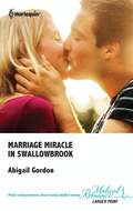
Marriage Miracle in Swallowbrook
by Abigail GordonThe husband she's never forgotten...Laura Armitage's heart broke the day she said goodbye to her husband, top oncologist Gabriel-but how could she stay in a marriage where she always came second to his career? Only, now Gabriel has joined Laura in the beautiful Lakeland village of Swallowbrook, and is determined to prove he's never stopped loving her...
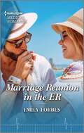
Marriage Reunion in the ER (Bondi Beach Medics #4)
by Emily ForbesWill the trauma surgeon be able to convince his estranged wife to start over again? Find out in the gripping final installment of the Bondi Beach Medics quartet by Emily Forbes.One chance……to win back his wife! Locking eyes over a patient isn&’t how ER doctor Lily expected to be reunited with her estranged husband, Otto. Two years ago, she lost their baby and fled to Sydney, unable to share her grief. And as far as Lily&’s concerned, their marriage is all but over. Yet trauma surgeon Otto is ready to fight for their relationship. Now he has three months to convince Lily they can make a fresh start…From Harlequin Medical: Life and love in the world of modern medicine. Bondi Beach MedicsBook 1: Rescuing the Paramedic's HeartBook 2: A Gift to Change His LifeBook 3: The Perfect Mother for His SonBook 4: Marriage Reunion in the ER
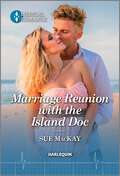
Marriage Reunion with the Island Doc
by Sue MacKayBeing stuck at a destination wedding with her ex-husband isn&’t the holiday she had planned! Find out if this could be their second chance in the latest Harlequin Medical Romance from Sue MacKay. A SECOND CHANCE IN PARADISE? At a destination wedding, nurse Alyssa is hoping for a stress-free break. But the best man is her ex-husband, island GP Leighton! Their whirlwind marriage collapsed as quickly as it began when Leighton suddenly withdrew from her. Alyssa never understood why, and now that they&’re stuck together for a fortnight, she wants to find out! Will her dream holiday end in closure…or a romantic reunion?From Harlequin Medical: Life and love in the world of modern medicine.
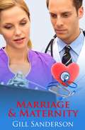
Marriage and Maternity
by Gill SandersonSister Angel Thwaite's neo-natal unit saves the life of an orphaned baby . To her horror she discovers that the baby's only relation is the baby's uncle, Dr Mike Gilmour has just started work in her hospital's cardiac unit. Mike is the surgeon who has to treat Angel's mother for a cardiac problem and eventually operate on her. He is also her ex-husband. Seven years ago they parted on bad terms.
Marriage and Maternity: A Story Of Lovers Reunited (Medical Romances #12)
by Gill SandersonAnother heartwarming medical romance from best-selling author Gill Sanderson! Perfect for fans of Mia Faye, Laura Scott, Helen Scott Taylor, Grey's Anatomy and ER.Readers LOVE Gill's gripping medical romances!'Outstanding writer and highly recommend this book!' 5* reader review'Another wonderful story I really enjoyed it' 5* reader review 'Well worth reading' 5* reader reviewSister Angel Thwaite's neo-natal unit saves the life of an orphaned baby. To her horror she discovers that the baby's uncle, Dr Mike Gilmour, has just started work in her hospital's cardiac unit. Mike is the surgeon who has to treat Angel's mother for a cardiac problem and eventually operate on her. However he is also her ex-husband, as they parted seven years ago, on bad terms.Can they put the past aside in order to give Angel's mother a chance for a future?Don't miss Gill Sanderson's enthralling medical romances, including the A Lakeland Practice and the Good, Bad and Ugly series.
Marriage and Maternity: A Story Of Lovers Reunited (Medical Romances Ser.)
by Gill SandersonAnother heartwarming medical romance from best-selling author Gill Sanderson! Perfect for fans of Mia Faye, Laura Scott, Helen Scott Taylor, Grey's Anatomy and ER.Readers LOVE Gill's gripping medical romances!'Outstanding writer and highly recommend this book!' 5* reader review'Another wonderful story I really enjoyed it' 5* reader review 'Well worth reading' 5* reader reviewSister Angel Thwaite's neo-natal unit saves the life of an orphaned baby. To her horror she discovers that the baby's uncle, Dr Mike Gilmour, has just started work in her hospital's cardiac unit. Mike is the surgeon who has to treat Angel's mother for a cardiac problem and eventually operate on her. However he is also her ex-husband, as they parted seven years ago, on bad terms.Can they put the past aside in order to give Angel's mother a chance for a future?Don't miss Gill Sanderson's enthralling medical romances, including the A Lakeland Practice and the Good, Bad and Ugly series.
Married for the Boss's Baby
by Susan CarlisleAn unexpected baby... When successful surgeon Grant Smythe's baby half sister is orphaned, Grant is determined to be there for little Lily-unlike his own father. A convenient proposal... But a challenge for custody means Grant needs a wife, too! New nanny Sara Marcum is the ideal candidate... A wife forever? It might be temporary, but soon warmhearted Sara completes more than just Grant's family. Can he convince his bride-for-now to become his forever wife?
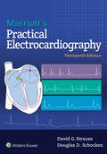
Marriott's Practical Electrocardiography
by David G Strauss Douglas SchockenProviding a solid foundation in current ECG technology as well as the newest diagnostic applications, Marriott’s Practical Electrocardiography, Thirteenth Edition, delivers the information you need to quickly improve your ECG interpretive skills. Authors Dr. David G. Strauss and Dr. Douglas D. Schocken offer medical students, residents and fellows a detailed explanation of the ECG, rhythm analysis, and how to rapidly and accurately interpret ECG results. Helpful diagrams, tables, animations, and videos, plus new ECG quizzes online, improve your understanding and skills.
Marrow of Tragedy: The Health Crisis of the American Civil War
by Margaret HumphreysSoldiers lay wounded or sick as both sides struggled to get them fit to return to battle.Winner, George Rosen Prize, American Association for the History of MedicineThe Civil War was the greatest health disaster the United States has ever experienced, killing more than a million Americans and leaving many others invalided or grieving. Poorly prepared to care for wounded and sick soldiers as the war began, Union and Confederate governments scrambled to provide doctoring and nursing, supplies, and shelter for those felled by warfare or disease. During the war soldiers suffered from measles, dysentery, and pneumonia and needed both preventive and curative food and medicine. Family members—especially women—and governments mounted organized support efforts, while army doctors learned to standardize medical thought and practice. Resources in the north helped return soldiers to battle, while Confederate soldiers suffered hunger and other privations and healed more slowly, when they healed at all.In telling the stories of soldiers, families, physicians, nurses, and administrators, historian Margaret Humphreys concludes that medical science was not as limited at the beginning of the war as has been portrayed. Medicine and public health clearly advanced during the war—and continued to do so after military hostilities ceased.
Marrying the Millionaire Doctor
by Alison RobertsGorgeous millionaire Dr. Vavunis has everything he needs in life--except a mother for his child Susie Jackson has come to Crocodile Creek's kids' camp to work--not to fall in love. Even if she did, it wouldn't be with the gorgeous but brooding Dr. Alex Vavunis. It's actually his daughter who captures Susie's heart--a girl desperately trying to reach out to her father. Susie realizes that single-father Alex needs to bond with his daughter, and she knows she can help. As she grows closer to the millionaire doctor, Susie sees she was wrong about him. Underneath the surface there is a perfect father--and husband--in the making!
Marrying the Playboy Doctor
by Laura IdingMoving to the small town of Cedar Bluff is a fresh start for paramedic Kylie Germaine and her young son, Ben. She's determined to forget the past and focus on her job--and raising Ben. But gorgeous emergency doctor Seth Taylor has other ideas. . . . Seth appreciates beautiful women, and he can't wait to get to know his new colleague better. But he soon discovers that, as a single mom, Kylie's priorities lie elsewhere. Still, for the first time Seth's smitten, and Cedar Bluff's most eligible bachelor finds himself wanting to put a ring on Kylie's finger and become a father to her little boy.
Mars and the Earthlings: A Realistic View on Mars Exploration and Settlement (Space and Society)
by Muriel Gargaud Kirsi Lehto Michel Viso Cyprien VerseuxIn an era of public Mars fascination, this book offers an objective presentation of the challenges of crewed Mars missions and discusses scenarios of Mars settlements under scientific, technical, social, economic , ethical and political aspects. With the aim to make the reader comprehend what is plausible and what is at stake, the book tries to clarify misconceptions and half-truths spreading rapidly in the public. The authors argue that approximations and misinformation should be countered for two main reasons. First, to avoid missing out on the benefits that Mars exploration may bring, including major scientific discoveries and an inspiring, federative human endeavor. Second, to remediate dangerous delusions – such as the idea that humanity could be transferred there should the Earth become inhabitable in the near term. In preparation for this book a group of European, world-renowned scientists from fields as diverse as astronomy, planetology, geology, biology, philosophy, or economics, as well as astronauts and science-fiction writers, was gathered to discuss Mars missions ranging from near-term robotic missions, all the way to large-scale settlements and even the feasibility of terraforming. For each, they draw arguments from their domains of expertise to discuss what is feasible and what is desirable. The result provides researchers with an objective review of the field, policy makers with a reference to make informed decisions, and the general public with a tool to form educated opinions.
Martin's Physical Pharmacy and Pharmaceutical Sciences
by Patrick J. SinkoConsistently revised and updated for more than 60 years to reflect the most current research and practice, Martin’s Physical Pharmacy and Pharmaceutical Sciences, 8th Edition, is the original and most comprehensive text available on the physical, chemical, and biological principles that underlie pharmacology and the pharmaceutical sciences. An ideal resource for PharmD and pharmacy students worldwide, teachers, researchers, or industrial pharmaceutical scientists, this 8th Edition has been thoroughly revised, enhanced, and reorganized to provide readers with a clear, consistent learning experience that puts essential principles and concepts in a practical, approachable context. Updated content reflects the latest developments and perspectives across the full spectrum of physical pharmacy and a new full-color design makes it easier than ever to discover, distinguish, and understand information—providing users the most robust support available for applying the elements of biology, physics, and chemistry in work or study.

Martini Fundamentals Of Anatomy And Physiology: Applications Manual
by William C. Ober Claire W. Garrison Ralph T. Hutchings Frederic Martini Kathleen WelchThis Applications Manual supplements your textbook and helps you apply the concepts of anatomy and physiology to your daily life. The information it contains will be useful to everyone and especially valuable for those planning a career as a health professional. Has a lot of photographs and illustrations to assist you.
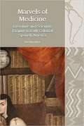
Marvels of Medicine: Literature and Scientific Enquiry in Early Colonial Spanish America (Liverpool Latin American Studies)
by Yarí Pérez MarínMarvels of Medicine makes a compelling case for including sixteenth century medical and surgical writing in the critical frameworks we now use to think about a genealogy of cultural expression in Latin America. Focusing on a small group of practitioners who differed in their levels of training, but who shared the common experience of having left Spain to join colonial societies in the making, this book analyses the paths their texts charted to attitudes and political positions that would come to characterize a criollo mode of enunciation. Unlike the accounts of first explorers, which sought to amaze audiences back in Europe with descriptions of strange and astonishing lands, these texts instead engaged the marvellous in an effort to supersede it, stressing the value of sensorial experience and of verifying information thorough repetition and demonstration. Vernacular medical writing became an unlikely early platform for a new form of regionally-anchored discourse that demanded participation in a global intellectual conversation, yet found itself increasingly relegated to the margins. In responding to that challenge, anatomical treatises, natural histories and surgical manuals exceeded the bounds set by earlier templates becoming rich, hybrid narratives that were as concerned with science as with portraying the lives and sensibilities of women and men in early colonial Mexico.
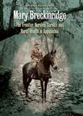
Mary Breckinridge
by Melanie Beals GoanIn 1925 Mary Breckinridge (1881-1965) founded the Frontier Nursing Service (FNS), a public health organization in eastern Kentucky providing nurses on horseback to reach families who otherwise would not receive health care. Through this public health organization, she introduced nurse-midwifery to the United States and created a highly successful, cost-effective model for rural health care delivery that has been replicated throughout the world.In this first comprehensive biography of the FNS founder, Melanie Beals Goan provides a revealing look at the challenges Breckinridge faced as she sought reform and the contradictions she embodied. Goan explores Breckinridge's perspective on gender roles, her charisma, her sense of obligation to live a life of service, her eccentricity, her religiosity, and her application of professionalized, science-based health care ideas. Highly intelligent and creative, Breckinridge also suffered from depression, was by modern standards racist, and fought progress as she aged--sometimes to the detriment of those she served.Breckinridge optimistically believed that she could change the world by providing health care to women and children. She ultimately changed just one corner of the world, but her experience continues to provide powerful lessons about the possibilities and the limitations of reform.
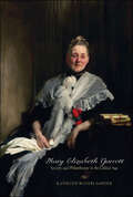
Mary Elizabeth Garrett: Society and Philanthropy in the Gilded Age
by Kathleen Waters SanderA captivating look at the remarkable life of this nineteenth-century suffragist, philanthropist, and reformer.Mary Elizabeth Garrett was one of the most influential philanthropists and women activists of the Gilded Age. With Mary's legacy all but forgotten, Kathleen Waters Sander recounts in impressive detail the life and times of this remarkable woman, through the turbulent years of the Civil War to the early twentieth century. At once a captivating biography of Garrett and an epic account of the rise of commerce, railroading, and women's rights, Sander's work reexamines the great social and political movements of the age.As the youngest child and only daughter of the B&O Railroad mogul John Work Garrett, Mary was bright and capable, well suited to become her father's heir apparent. But social convention prohibited her from following in his footsteps, a source of great frustration for the brilliant and strong-willed woman. Mary turned her attention instead to promoting women's rights, using her status and massive wealth to advance her uncompromising vision for women's place in the expanding United States. She contributed the endowment to establish the Johns Hopkins School of Medicine with two unprecedented conditions: that women be admitted on the same terms as men and that the school be graduate level, thereby forcing revolutionary policy changes at the male-run institution. Believing that advanced education was the key to women's betterment, she helped found and sustain the prestigious girls' preparatory school in Baltimore, the Bryn Mawr School. Her philanthropic gifts to Bryn Mawr College helped transform the modest Quaker school into a renowned women's college. Mary was also a great supporter of women's suffrage, working tirelessly to gain equal rights for women.Suffragist, friend of charitable causes, and champion of women's education, Mary Elizabeth Garrett both improved the status of women and ushered in modern standards of American medicine and philanthropy. Sander's thoughtful and informed study of this pioneering philanthropist is the first to recognize Garrett and her monumental contributions to equality in America.
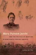
Mary Putnam Jacobi and the Politics of Medicine in Nineteenth-Century America
by Carla BittelIn the late nineteenth century, as Americans debated the "woman question," a battle over the meaning of biology arose in the medical profession. Some medical men claimed that women were naturally weak, that education would make them physically ill, and that women physicians endangered the profession. Mary Putnam Jacobi (1842-1906), a physician from New York, worked to prove them wrong and argued that social restrictions, not biology, threatened female health. Mary Putnam Jacobi and the Politics of Medicine in Nineteenth-Century America is the first full-length biography of Mary Putnam Jacobi, the most significant woman physician of her era and an outspoken advocate for women's rights. Jacobi rose to national prominence in the 1870s and went on to practice medicine, teach, and conduct research for over three decades. She campaigned for co-education, professional opportunities, labor reform, and suffrage--the most important women's rights issues of her day. Downplaying gender differences, she used the laboratory to prove that women were biologically capable of working, learning, and voting. Science, she believed, held the key to promoting and producing gender equality. Carla Bittel's biography of Jacobi offers a piercing view of the role of science in nineteenth-century women's rights movements and provides historical perspective on continuing debates about gender and science today.
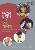
Mary Sheridan's From Birth to Five Years: Children's Developmental Progress
by Helen Cockerill Ajay SharmaFrom Birth to Five Years, based on the pioneering work of Mary Sheridan, is widely regarded as the go-to reference for health, education and social care professionals, or anyone concerned with the developmental progress of pre-school children. In this new fourth edition, the text has been developed to further align it with current child development philosophies and practices, and to support the wider group of professionals that are now required to take steps for promoting children’s development as part of their assessment and management plans. This book aims to improve the clinical management of children with developmental disorders, through providing the full range of developmental attainments, methods of observation, and advice about when to seek help. Features of this completely revised edition include: For students and tutors – information on theoretical aspects of development, with further reading suggestions and references including the most recent international studies in the field A new section on the development of attention and self-regulation Contemporary case studies with guidance on when to raise concerns for students and teachers Discussion points to stimulate class debate To complement this book, a new companion volume, From Birth to Five Years: Practical Developmental Examination, offers a step-by-step ‘how to’ guide, including guidance on enquiry and observation, how to chart typical and atypical patterns, and ‘red flags’ for recognising significant delay or abnormality. To consolidate and expand on the practical and theoretical information across both books, a new companion website is available at www.routledge.com/cw/sharma, which includes the following additional learning material: An interactive timeline of the key developmental domains Introductions to theory with links to further reading Research summaries Video clips demonstrating practical assessment skills
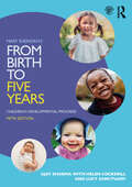
Mary Sheridan's From Birth to Five Years: Children's Developmental Progress
by Helen Cockerill Ajay Sharma Lucy SanctuaryThis new edition of a classic text is the go-to reference for anyone concerned with the developmental progress of pre-school children. It provides the knowledge required for understanding children’s developmental progress with age and within each developmental domain. Including new sections on atypical development for each of the core domains of development and additional material on the development of attention and self-regulation, this fifth edition integrates findings from the latest research throughout. An updated companion website is available at www.routledge.com/cw/sharma, which includes the following additional learning material: an interactive timeline of the key developmental domains; introductions to theory with links to further reading; research summaries; video clips demonstrating practical assessment skills; downloadable resources including pictures to support examination of verbal and non-verbal development, and tips to facilitate and promote development. Fully aligned with current child development philosophies and practices, Mary Sheridan’s From Birth to Five Years: Children's Developmental Progress is designed to support the wider group of practitioners – including those from health professions, social work and early years – that are now required to take steps for promoting children’s development as part of their assessment and management plans.
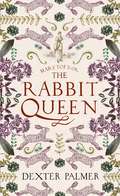
Mary Toft; or, The Rabbit Queen: A Novel
by Dexter Palmer'Palmer spins a cracking tale that, despite its disconcerting subject, is piquantly cheerful and compassionate . . . With empathy and imagination, Palmer explores the master/apprentice relationship, first love and first rivalry, spite and kindness: conjuring a world to raise a wry smile' New York Times---------------------------------------------------------------------------------A stunning, powerfully evocative new novel based on a true story - in 1726 in the small town of Godalming, England, a young woman confounds the medical community by giving birth to dead rabbits.Surgeon John Howard is a rational man. His apprentice Zachary knows John is reluctant to believe anything that purports to exist outside the realm of logic. But even John cannot explain how or why Mary Toft, the wife of a local farmer, manages to give birth to a dead rabbit. When this singular event becomes a regular occurrence, John realizes that nothing in his experience as a village physician has prepared him to deal with a situation as disturbing as this. He writes to several preeminent surgeons in London, three of whom quickly arrive in the small town of Godalming ready to observe and opine. When Mary's plight reaches the attention of King George, Mary and her doctors are summoned to London, where Zachary experiences for the first time a world apart from his small-town existence, and is exposed to some of the darkest corners of the human soul. All the while, Mary lies in bed, waiting for another birth, as doubts begin to blossom among the surgeons and a growing group of onlookers grow impatient for another miracle . . .