- Table View
- List View
Olga: A Novel
by Prof Bernhard SchlinkA #1 INTERNATIONAL BESTSELLER'Bernhard Schlink speaks straight to the heart' New York Times'Brilliant... A tale of love and loss in 20th century Germany' Evening Standard'A cleverly-constructed tale of cross-class romance' Mail on Sunday'A poignant portrait of a woman out of step with her time' Observer Olga is an orphan raised by her grandmother in a Prussian village around the turn of the 20th century. Smart and precocious, she fights against the prejudices of the time to find her place in a world that sees her as second-best.When she falls in love with Herbert, a local aristocrat obsessed with the era's dreams of power, glory and greatness, her life is irremediably changed.Theirs is a love against all odds, entwined with the twisting paths of German history, leading us from the late 19th to the early 21st century, from Germany to Africa and the Arctic, from the Baltic Sea to the German south-west.This is the story of that love, of Olga's devotion to a restless man - told in thought, letters and in a fateful moment of great rebellion.
Oligodendrocytes: Methods and Protocols (Methods in Molecular Biology #1936)
by David A. Lyons Linde KegelThis volume looks at the study of oligodendrocytes through in vitro and in vivo techniques, multiple model organisms, using approaches that bridge scales from molecular through system. Chapters in this book cover topics such as fundamental molecular analyses of oligodendrocytes and myelin; in vitro, ex vivo, and in vivo molecular-cellular-electrophysiology-based techniques; oligodendrocyte formation, homeostasis, and disruption in zebrafish and Xenopus; and parallel system-level imaging of animal and human models. Written in the highly successful Methods in Molecular Biology series format, chapters include introductions to their respective topics, lists of the necessary materials and reagents, step-by-step, readily reproducible laboratory protocols, and tips on troubleshooting and avoiding known pitfalls.Authoritative and thorough, Oligodendrocytes: Methods and Protocols is a valuable reference guide that highlights the expansive and fast-paced nature of research into oligodendrocyte biology underlying health and function.
Oligonucleotide Synthesis
by Piet HerdewijnA collection of powerful new techniques for oligonucleotide synthesis and for the use of modified oligonucleotides in biotechnology. Among the protocol highlights are a novel two-step process that yields a high purity, less costly, DNA, the synthesis of phosphorothioates using new sulfur transfer agents, the synthesis of LNA, peptide conjugation methods to improve cellular delivery and cell-specific targeting, and triple helix formation. The applications include using molecular beacons to monitor the PCR amplification process, nuclease footprinting to study the sequence-selective binding of small molecules of DNA, nucleic acid libraries, and the use of small interference RNA (siRNA) as an inhibitor of gene expression.
Oligonucleotide-Based Drugs and Therapeutics: Preclinical and Clinical Considerations for Development
by Nicolay Ferrari Rosanne SeguinA comprehensive review of contemporary antisense oligonucleotides drugs and therapeutic principles, methods, applications, and research Oligonucleotide-based drugs, in particular antisense oligonucleotides, are part of a growing number of pharmaceutical and biotech programs progressing to treat a wide range of indications including cancer, cardiovascular, neurodegenerative, neuromuscular, and respiratory diseases, as well as other severe and rare diseases. Reviewing fundamentals and offering guidelines for drug discovery and development, this book is a practical guide covering all key aspects of this increasingly popular area of pharmacology and biotech and pharma research, from the basic science behind antisense oligonucleotides chemistry, toxicology, manufacturing, to safety assessments, the design of therapeutic protocols, to clinical experience. Antisense oligonucleotides are single strands of DNA or RNA that are complementary to a chosen sequence. While the idea of antisense oligonucleotides to target single genes dates back to the 1970's, most advances have taken place in recent years. The increasing number of antisense oligonucleotide programs in clinical development is a testament to the progress and understanding of pharmacologic, pharmacokinetic, and toxicologic properties as well as improvement in the delivery of oligonucleotides. This valuable book reviews the fundamentals of oligonucleotides, with a focus on antisense oligonucleotide drugs, and reports on the latest research underway worldwide. • Helps readers understand antisense molecules and their targets, biochemistry, and toxicity mechanisms, roles in disease, and applications for safety and therapeutics • Examines the principles, practices, and tools for scientists in both pre-clinical and clinical settings and how to apply them to antisense oligonucleotides • Provides guidelines for scientists in drug design and discovery to help improve efficiency, assessment, and the success of drug candidates • Includes interdisciplinary perspectives, from academia, industry, regulatory and from the fields of pharmacology, toxicology, biology, and medicinal chemistry Oligonucleotide-Based Drugs and Therapeutics belongs on the reference shelves of chemists, pharmaceutical scientists, chemical biologists, toxicologists and other scientists working in the pharmaceutical and biotechnology industries. It will also be a valuable resource for regulatory specialists and safety assessment professionals and an important reference for academic researchers and post-graduates interested in therapeutics, antisense therapy, and oligonucleotides.
Oligonucleotide-Based Therapies: Methods and Protocols (Methods in Molecular Biology #2036)
by Olof Gissberg Rula Zain Karin E. LundinThis book provides a collection of comprehensive, up-to-date, and broadly applicable guides to the research and development fields of oligonucleotide (ON) therapeutics. Covering topics from the study of antisense and anti-gene effects to oligonucleotides in the context of drug discovery and development, the volume explores a wide-ranging and useful spectrum of methods and protocols needed to take full advantage of therapeutic applications involving ONs. Written for the highly successful Methods in Molecular Biology series, chapters include introductions to their respective topics, lists of the necessary materials and reagents, step-by-step, readily reproducible laboratory protocols, and tips on troubleshooting and avoiding known pitfalls. Authoritative and practical, Oligonucleotide-Based Therapies: Methods and Protocols aims to be a great aid in the laboratory as well as an ideal reference guide when designing antisense and anti-gene oligonucleotides for therapeutic applications.
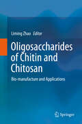
Oligosaccharides of Chitin and Chitosan: Bio-manufacture and Applications
by Liming ZhaoThe book provides an overview of bio-manufacturing techniques for the production, purification, characterization and modification of chito/chitin oligosaccharides and their monomers. In addition, it explores potential applications in the food, biomedical and agricultural industry on the basis of their bioactivities and biomaterial properties. Lastly, it shares a range of cutting-edge insights to help solve problems in industrial processes and promote further academic investigation. Given its scope, it offers a valuable resource for researchers and graduate students in the fields of bioengineering, food science, biochemistry, etc.
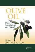
Olive Oil: Minor Constituents and Health
by Dimitrios BoskouEpidemiological studies indicate that the consumption of natural antioxidants from such plant-derived sources as olive oil produces beneficial health effects. Olive Oil: Minor Constituents and Health provides a balanced understanding of the pharmacological properties of phenols and other bioactive ingredients in the composition of olive oil. It dis
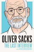
Oliver Sacks: The Last Interview
by Oliver SacksAn extraordinary collection of interviews with the beloved doctor and author, whose research and books inspired generations of readers.Oliver Sacks--called "the poet laureate of medicine" by the New York Times--illuminated the mysteries of the brain for a wide audience in a series of richly acclaimed books, including Awakenings and The Man Who Mistook His Wife for a Hat, and numerous The New Yorker articles. In this collection of interviews, Sacks is at his most candid and disarming, rich with insights about his life and work. Any reader of Oliver Sacks will find in this book an entirely new way of looking at a brilliant writer.
Olivia: My Life of Exile in Kalaupapa
by Olivia Robello BreithaOlivia Robello Breitha was diagnosed with Hansen's disease (leprosy) when she was a young woman in the 1930s. At the time, there was no treatment for the disease and in Hawaii many people with leprosy were exiled to Kalaupapa, a community on the island of Molokai. Breitha, who had a 6th grade education, wrote her powerful story about living with the disease. At times they were treated inhumanely, mostly by doctors and medical professionals, and Breitha was unafraid to fight for patient rights and reducing stigma.
Omega Balance: Nutritional Power for a Happier, Healthier Life (A Johns Hopkins Press Health Book)
by Anthony John HulbertLearn how to live a happier and healthier life by finding the right balance of omega fatty acids in your diet.Omega-3 and omega-6 fatty acids are essential in the human diet. In Omega Balance, noted scientist Anthony J. Hulbert explains how the balance between these fatty acids in the human food chain has changed over the last half-century and the very serious negative health impacts this imbalance has created. An imbalance of these omega fats contributes to increased rates of obesity, diabetes, cardiovascular diseases, allergies, asthma, as well as cancer and a variety of other inflammatory diseases. Omega balance is also important for normal brain function, and an imbalance in these fatty acids is associated with depression, mood disorders, and neurodegenerative diseases. Hulbert provides extensive information on the omega balance of different foods and discusses fascinating details about human evolution, dietary changes throughout history, the effect of diet on human development and physiological processes, and more. He investigates the paleo diet of our ancestors and describes the dramatic changes that have accompanied the increased ultra-processing of modern foods. Omega Balance is an essential guide to understanding a significant problem in our modern food chain and will make us rethink the food we eat.
Omega-3 Fatty Acids
by Mahabaleshwar V. Hegde Anand Arvind Zanwar Sharad P. AdekarThis volume argues for the importance of essential nutrients in our diet. Over the last two decades there has been an explosion of research on the relationship of Omega-3 fatty acids and the importance of antioxidants to human health. Expert authors discuss the importance of a diet rich in Omega-3 Fatty acids for successful human growth and development and for the prevention of disease. Chapters highlight their contribution to the prevention and amelioration of a wide range of conditions such as heart disease, diabetes, arthritis, cancer, obesity, mental health and bone health. An indispensable text designed for nutritionists, dietitians, clinicians and health related professionals, Omega-3 Fatty Acids: Keys to Nutritional Health presents a comprehensive assessment of the current knowledge about the nutritional effects of Omega-3 fatty acids and their delivery in foods.
Omega-3 Fatty Acids and the DHA Principle
by Raymond C. Valentine David L. ValentineThe physical-chemical properties of the omega-3 fatty acid DHA (docosahexaenoic acid) enable it to facilitate rapid biochemical processes in the membrane. This effect has numerous benefits, including those involved in the growth of bacteria, rapid energy generation, human vision, brain impulse, and photosynthesis, to name a few. Yet DHA also carrie
Omega-3 Fatty Acids in Health and Disease
by Robert S. LeesA report from research in the MIT Sea Grant College Program. Discusses the relationship between particular fatty acids found only in fish oil, and human health. Presents and evaluates information on the health effects of dietary fats generally; evidence that fish oil consumption affects the incidenc
Omega-3 Fatty Acids: Keys to Nutritional Health and Disease
by Mahabaleshwar V. Hegde Anand Arvind Zanwar Sharad P. AdekarThis book argues for the importance of omega-3 fatty acids in our diet. Omega-3 fatty acids are a must in our daily diet, as the human body cannot synthesize it. The human body is crippled in evolution; we are deprived of the genes that are needed to synthesize these vital molecules. Except for regular fish eaters, the majority of the human population does not get adequate omega-3 fatty acid in their food. Fatty acids provide a structural framework for cells, tissues, and organs, as well as the building blocks for several bioactive ingredients, and they provide a wide range of benefits from general improvements in health to protection against inflammation and disease. Omega-3 Fatty Acids discusses various sources of omega-3 fatty acid, health implications of omega-3 fatty acid intake, and remedial measures that can improve diet for those lacking in fatty acids. The book opens with a discussion of various sources of omega-3 fatty acids, such as flaxseed, milk, eggs, and marine algae. Following this, is a detailed discussion of the effect omega-3 intake has on different conditions, like pregnancy, psoriasis, aging disorders, cardiovascular events, obesity, and non-communicable diseases, such as diabetes and Alzheimer’s. This much-expanded edition includes new chapters on topics such as the linoleic-to-linolenic dietary intake ratio, the role of omega-3 fatty acids in eye health, the effects of omega-3 fatty acids on metabolic syndrome and fatty liver disease, and the influence of omega-3 fatty acids on bone turnover and energy metabolism. An indispensable text designed for nutritionists, dietitians, clinicians and health-related professionals, Omega-3 Fatty Acids presents a comprehensive assessment of the current knowledge about the nutritional effects of omega-3 fatty acids and their delivery in foods.
Omega-6/3 Fatty Acids
by Ronald Ross Watson Sherma Zibadi Fabien De MeesterOver the last several years developing human research suggests that a component of omega-3 fatty acids, long chain ones, contribute particularly to health benefits. Omega-6/3 Fatty Acids: Functions, Sustainability Strategies and Perspectives focuses on developing information on this newly recognized key component. This volume uniquely, and for the first time, focuses on sustainability of natural sources of omega-3 fatty acids variants including long chain ones, and on ways to increase their use and availability to reduce major diseases. The authors review cardiovascular disease, neurological changes and mental health and other diseases like diabetes where long chain omega-3 fatty acids play protective roles from recent human trials. Each chapter evaluates developing information on the possible mechanistic role of long chain omega-3 fatty acids. After showing their requirement and involvement in health promotion there are reviews of various sources and ways to protect and promote them. Authors provide support for the benefits and sources of long chain omega-3 fatty acids and their increased dietary intake that reduce various physical and mental illnesses. Omega-6/3 Fatty Acids: Functions, Sustainability and Perspectives is a unique and important new volume that provides the latest data and reviews to physicians who need to assess serum omega-6/3 and fatty acids to help diagnose risks and change diets and to inform industry and the scientific community with reviews of research for actions including new studies and therapies.
Omgaan met een scheiding
by J.P. van de Ven BVEr gaan in Nederland elke dag driehonderd stellen uit elkaar (dit zijn op jaarbasis 219. 000 mensen). Zij raken veelal verwikkeld in het afhandelen van allerhande juridische en praktische zaken. Maar over gevoelens zoals verwarring, angst, woede, wantrouwen, jaloezie, verdriet, opluchting, schuld en schaamte die met een scheiding gepaard gaan, en over hoe je daarmee omgaat is weinig informatie voorhanden. Het boek omgaan met een scheiding voorziet hierin. Het behandelt de gevolgen van een scheiding op emotioneel, mentaal en sociaal gebied.
Omgaan met hoogtevrees.
by S. J. Swaen Vogelbescheming Nederland W. A. Sterk IpzoHoogtevrees is een van de bekendste vormen van angst. De klachten treden niet alleen op grote hoogtes op. Sommige mensen durven bijvoorbeeld niet in diep helder water te zwemmen of hebben moeite om naar een kerktoren op te kijken. Iedereen heeft in zekere mate last van hoogtevrees. Hoogtes kunnen gevaarlijk zijn en het is dan ook logisch om op grote hoogte op je hoede te zijn. Maar wanneer de angst uit de hand loopt en je bang wordt in situaties die geen angst hoeven op te wekken, dan belemmert de hoogtevrees je dagelijks leven. Mensen met hoogtevrees gaan immers steeds meer situaties uit de weg waarin ze met hoogteverschillen te maken kunnen krijgen. Overmatige hoogtevrees is daarom een vervelende klacht, maar doorgaans goed te verhelpen. Dit boek laat zien dat iedereen met wat motivatie, begeleiding en tijdsinvestering zijn hoogtevrees onder controle kan krijgen. Hierbij wordt uitgegaan van de principes van de gedragstherapie. Door de stapsgewijze beschrijving van de gedragstherapeutische weg met duidelijke tips en opdrachten is Omgaan met hoogtevrees een waardevol zelfhulpboek. De heldere beschrijvingen van andere therapieën met een indicatie van de effectiviteit ervan maken het ook een handige wegwijzer voor wie liever een beroep doet op de hulpverlening. Omgaan met hoogtevrees verschijnt in de reeks Van A tot ggZ.
Omgaan met onverklaarde lichamelijke klachten
by A. Blok J. HoutveenInformatief boek voor mensen met onverklaarde lichamelijke klachten.
Omgaan met slaapproblemen
by C. Kluft G. KerkhofBijna een derde van de Nederlandse bevolking heeft regelmatig of vaak last van slapeloosheid. Slapeloosheid houdt in dat je moeite hebt om in slaap te komen en lang wakker ligt. Ook kan het voorkomen dat je in de loop van de nacht meerdere malen wakker wordt. We spreken van een slaapprobleem wanneer de slapeloosheid niet vanzelf overgaat en klachten oplevert in het functioneren overdag. Wat zijn de kenmerken van chronische slapeloosheid? Wat zijn veelvoorkomende oorzaken? En welke behandelmethoden kunnen toegepast worden? De auteurs geven antwoord op deze vragen, en laten zien wat cognitieve gedragstherapie voor insomnie inhoudt. Zij nemen het slaapgedrag onder de loep en bespreken mogelijke aanpassingen, met het doel om de kans op goed in- en doorslapen te vergroten. Omgaan met slapeloosheid is een nieuw deel in de reeks Van A tot ggZ. Deze reeks is geschikt voor iedereen die te maken heeft met psychische of sociale problemen, zowel voor de betrokkenen zelf als voor de omgeving. De boeken worden veelvuldig gebruikt door (huis)artsen, psychologen, psychiaters, psychotherapeuten, hulpverleners, maatschappelijk werkers en andere professionals in de gezondheidszorg. Niet alleen om zelf meer inzicht te krijgen in de verschillende problemen en hoe mensen deze beleven, maar vooral om hun cliënten voor te lichten.
Omgaan met stress en burnout
by Bart Verkuil Arnold Van EmmerikStress is vaak erg nuttig. Wie kent niet de gezonde spanning die vooraf gaat aan het leveren van een belangrijke prestatie? Door de stress presteer je vaak nét iets beter. Diezelfde stress zorgt er ook voor dat je in gevaarlijke situaties de kracht vindt om in actie te komen: om te vluchten of om je te verdedigen. Stress speelt dus een duidelijke rol in ons vermogen te overleven. Maar soms lukt het niet om stress om te zetten in actie. De spanning maakt dan geen plaats voor voldoening of opluchting, maar blijft langdurig hangen. Dat is vervelend en kan ook gevaarlijk zijn. Die ongezonde stress kost namelijk veel energie en kan uiteindelijk leiden tot overspannenheid, burnout en zelfs depressie. Omgaan met stress en burnout laat zien hoe je in zo'n situatie terecht kunt komen en leert je uit te zoeken welke activiteiten jou energie kosten en welke je juist energie opleveren. Je leert prioriteiten te stellen en voor jezelf op te komen. De auteurs beschrijven hoe je met je gedachten je gevoel beïnvloedt en geven diverse technieken waarmee je kunt ontspannen en de controle leert houden over je gedachten. De praktische tips en oefeningen uit dit boek helpen je zo om ongezonde stress te verminderen en je leven (weer) in balans te brengen.
Omics Applications for Systems Biology (Advances in Experimental Medicine and Biology #1102)
by Wan Mohd Aizat Hoe-Han Goh Syarul Nataqain BaharumThis book explains omics at the most basic level, including how this new concept can be properly utilized in molecular and systems biology research. Most reviews and books on this topic have mainly focused on the technicalities and complexity of each omics’ platform, impeding readers to wholly understand its fundamentals and applications. This book tackles such gap and will be most beneficial to novice in this area, university students and even researchers. Basic workflow and practical guidance in each omics are also described, such that scientists can properly design their experimentation effectively. Furthermore, how each omics platform has been conducted in our institute (INBIOSIS) is also detailed, a comprehensive example on this topic to further enhance readers’ understanding. The contributors of each chapter have utilized the platforms in various manner within their own research and beyond. The contributors have also been interactively integrated and combined these different omics approaches in their research, being able to systematically write each chapter with the conscious knowledge of other inter-relating topics of omics. The potential readers and audience of this book can come from undergraduate and postgraduate students who wish to extend their comprehension in the topics of molecular biology and big data analysis using omics platforms. Furthermore, researchers and scientists whom may have expertise in basic molecular biology can extend their experimentation using the omics technologies and workflow outlined in this book, benefiting their research in the long run.
Omics Approaches for Sugarcane Crop Improvement
by Rajarshi Kumar GaurIn this book, the information encompasses various researchable biotechnology aspects of sugarcane, its genomic structure, diversity, comparative and structural genomics, data mining, etc. This book explores both the theoretical and practical aspects of sugarcane crops, focusing on innovative processes. This book argues in favor of developing an integrated research and development system to strengthen the research and development capabilities of all the areas of sugarcane. Further, it covers the recent trends of sugarcane biotechnology, especially in the next-generation sequencing (NGS) era. This book will be very useful for professors and scientists who are working in the area of sugarcane crops by using molecular biology and bioinformatics. It is also useful for students to use as a reference for their classes or thesis projects. Key features: • Discusses an integral part of molecular biology and pivotal tools for molecular breeding; enables breeders to design cost-effective and efficient breeding strategies for sugarcane • Discusses the harnessing genomics technologies for genetic engineering and pathogen characterization and diagnosis of sugarcane • Provides new examples and problems, added where needed • Provides insight from contributors drawn from around the globe
Omics Approaches in Breast Cancer
by Debmalya BarhBreast cancer is the most common cancer in females that accounts for highest cancer specific deaths worldwide. In the last few decades research has proven that breast cancer can be treated if diagnosed at early stages and proper therapeutic strategy is adopted. Omics-based recent approaches have unveiled the molecular mechanism behind the breast tumorigenesis and aid in identification of next-generation molecular markers for early diagnosis, prognosis and even the effective targeted therapy. Significant development has taken place in the field of omics in breast cancer in the last decade. The most promising omics approaches and their outcomes in breast cancer have been presented in this book for the first time. The book covers omics technologies and budding fields such as breast cancer miRNA, lipidomics, epigenomics, proteomics, nutrigenomics, stem cell, pharmacogenomics and personalized medicine and many more along with conventional topics such as breast cancer management etc. It is a research-based reference book useful for clinician-scientists, researchers, geneticists and health care industries involved in various aspects of breast cancer. The book will also be useful for students of biomedicine, pathology and pharmacy.
Omics Approaches in Veterinary Parasitology: Diagnosis, Biomarkers, and Drug Development
by Muhammad Sohail Sajid and Hafiz Muhammad RizwanOmics Approaches in Veterinary Parasitology: Diagnosis, Biomarkers, and Drug Development explores applications of omics approaches for diagnosis, biomarker discovery, and drug development against parasites of veterinary importance. It presents the fundamental principles of parasite biology and their complex physiological processes. The chapters review key aspects such as parasite life cycles, host-parasite interactions, and the molecular mechanisms that underlie parasitic diseases. The subsequent chapters delve into the principles and applications of genomics, transcriptomics, proteomics, and metabolomics in understanding parasites at a molecular level. The use of next-generation sequencing, PCR-based assays, and metagenomics in identifying and characterizing parasites for accurate and efficient diagnosis are also covered in detail. Toward the end, the book focuses on target identification, drug repurposing, and the optimization of drug efficacy while minimizing drug resistance using omics data. The book is useful for researchers, students, and professionals in the field of veterinary parasitology.
Omics Approaches to Understanding Muscle Biology (Methods in Physiology)
by Jatin George Burniston Yi-Wen ChenThis book is a collection of principles and current practices in omics research, applied to skeletal muscle physiology and disorders. The various sections are categorized according to the level of biological organization, namely, genomics (DNA), transcriptomics (RNA), proteomics (protein), and metabolomics (metabolite). With skeletal muscle as the unifying theme, and featuring contributions from leading experts in this traditional field of research, it highlights the importance of skeletal muscle tissue in human development, health and successful ageing. It also discusses other fascinating topics like developmental biology, muscular dystrophies, exercise, insulin resistance and atrophy due to disuse, ageing or other muscle diseases, conveying the vast opportunities for generating new hypotheses as well as testing existing hypotheses by combining high-throughput techniques with proper experiment designs, bioinformatics and statistical analyses. Presenting the latest research techniques, this book is a valuable resource for the physiology community, particularly researchers and grad students who want to explore the new opportunities for omics technologies in basic physiology research.

