- Table View
- List View
PET in the Evaluation of Alzheimer's Disease and Related Disorders
by Dan SilvermanThis practical guide contains all the science and clinical information needed to help the reader make informed decisions about the applications of brain PET to improve the care of patients with symptoms of Alzheimer's disease and related disorders. Beginning with an introduction to goals and limitations of the clinical evaluation of dementia, Dr. Dan Silverman and his distinguished group of contributors cover the role of functional imaging, clinical interpretation of brain FDG-PET scans, FDG-PET in the evaluation of early dementia, microstructural imaging of neurodegenerative changes, amyloid imaging, imaging non-neurodegenerative causes of cognitive impairment, tools for assisting PET scan interpretation in the clinical setting, and PET evaluation of central motor disorders. There is also a complete collection of clinical cases that practitioners are likely to encounter in daily practice. Enhanced with numerous PET brain scans, this book fills a rapidly growing need in the medical imaging field.
PET-CT Beyond FDG
by Mohsen Farsad Stefano Fanti Luigi MansiAlthough [18F]fluorodeoxyglucose (FDG) generally shows an excellent performance as a cancer-imaging agent when using PET-CT, there are some settings in which other radiopharmaceuticals offer advantages. Such non-FDG tracers are now gaining widespread acceptance not only in research but also in clinical practice. This atlas, including about 500 high-quality images, is a user-friendly guide to PET-CT imaging beyond FDG. A wide range of tracers is covered, such as 18F- and 11C-choline, 11C-methionine, 18F-ethyl-L-tyrosine, 68Ga-DOTA-NOC, 11C-acetate, 11C-thymidine, and 18F-DOPA. Throughout, the emphasis is on image interpretation, with guidance on the recognition of normal, benign, and malignant uptake and clear instruction on learning points and pitfalls. This atlas is designed to serve as a reference text for both nuclear physicians and radiologists, and will also be of great benefit to radiographers, technologists, and nuclear medicine and radiology residents.
PET-CT and PET-MRI in Neurology
by Luigi Mansi Andrea CiarmielloUsing SWOT analysis, this book examines in detail the strengths and weaknesses of the hybrid modalities PET-CT and PET-MRI for imaging of the central nervous system, comparing their merits and evaluating their advantages over the stand-alone modalities. The aim is to employ a truly systematic approach in order to define the potential clinical benefit of these modalities and to identify shortcomings, opportunities, and threats. Clinical application of the modalities is explored in a range of conditions, including dementia and related disorders, movement disorders, psychiatric disorders, cerebrovascular disease, infection/inflammation, brain tumors, and pediatric neurologic disorders. In addition, the basics of hybrid imaging are addressed, covering physics, instrumentation, data analysis and quantitation, radiopharmaceuticals, and contrast media. PET-CT and PET-MRI in Neurology, written by experts from Europe and the United States, will be essential reading for imaging specialists and of value for neurologists, psychiatrists, neurosurgeons, and pediatricians.
PET-CT and PET-MRI in Oncology
by Patrick Peller Rathan Subramaniam Ali GuermaziOver the past decade, PET-CT has achieved great success owing to its ability to simultaneously image structure and function, and show how the two are related. More recently, PET-MRI has also been developed, and it represents an exciting novel option that promises to have applications in oncology as well as neurology. The first part of this book discusses the basics of these dual-modality techniques, including the scanners themselves, radiotracers, scan performance, quantitation, and scan interpretation. As a result, the reader will learn how to perform the techniques to maximum benefit. The second part of the book then presents in detail the PET-CT and PET-MRI findings in cancers of the different body systems. The final two chapters address the use of PET/CT in radiotherapy planning and examine areas of controversy. The authors are world-renowned experts from North America, Europe, and Australia, and the lucid text is complemented by numerous high-quality illustrations.
PET-CT in Infection and Inflammation
by Sikandar ShaikhThis book covers all aspects of PET-CT including newer applications involving infection and inflammation and basic information on PET-CT new tracers. It explains the approach, protocols and miscellaneous applications about various infections and inflammation that helps in its early and better diagnosis in multiorgan and multisystem involvements in routine clinical practice.Chapters provide systematic approach to the diagnosis of various infections such as bacterial, viral, tuberculosis, inflammatory pathologies and overlap in coexisting tumors and superadded/associated infections. This book offers assistance to medical practitioners in understanding various advantages of PET-CT in single scan as compared to different modalities for diagnosis as well as oncologists dealing with concurrent malignant and non-malignant conditions, students, and interns.
PET-CT: Rare Findings and Diseases
by Stefano Fanti Cristina NanniPET-CT is increasingly being employed in the diagnosis of both oncological and non-oncological patients, yet nuclear medicine physicians may have only limited practical experience of rare diseases and may experience difficulty in recognizing and interpreting rare findings. This unique atlas documents a large number of clinical cases that will help practitioners to identify findings and diseases that, though rare, are sufficiently frequent to be encountered in routine practice. Two types of cases are presented: patients evaluated for rare diseases and patients evaluated for standard diseases in whom atypical PET findings were detected. Each reported case includes a brief description of the clinical history, representative color PET-CT images obtained using FDG or other tracers, and a short explanation of the disease and findings. This atlas will enable practitioners to make conclusive reports of PET-CT scans that would otherwise have been inconclusive.
PET/CT Imaging: Basics and Practice (Clinicians’ Guides to Radionuclide Hybrid Imaging)
by Kanhaiyalal Agrawal Annah Skillen Abdulredha Esmail Sharjeel UsmaniThe aim of this book is to provide concise information and quick reference on the basics and practice of PET/CT for beginners. The chapters are written by Nuclear Medicine experts from different countries with enormous experience in PET/CT practice. Starting with the basics of PET/CT describing physics and the use of radiopharmaceuticals in PET/CT, the book explores the principle of PET/CT in radiotherapy planning. The last five chapters explore normal variation, pitfalls and artefacts commonly seen with various routinely used PET radiotracers. The text is enriched by tables and highlighted clinical cases for better understanding. This book will be of interest mostly to nuclear medicine physicians and radiologists, but it may be appealing also to a wider medical community including oncologists and radiotherapists.
PET/CT and PET/MR in Melanoma and Sarcoma
by Amir H. KhandaniThis is a comprehensive guide for patient preparation, image acquisition, and image interpretation for PET/CT and PET/MR, specifically relevant to melanoma and sarcoma. Imaging specialists and referring physicians are often not as intimately aware of the particulars of PET imaging in management of patients with melanoma and sarcoma and how it could affect their treatment. This book fills that gap by presenting comprehensive information on melanoma, sarcoma, and the role of PET imaging in their diagnosis and management. The book begins by covering the basics of imaging for practicing physicians and trainees. Expert authors then further cover the biological concepts of melanoma and sarcoma and how they relate to imaging, particularly PET, the oncologist’s perspective, and the surgeon’s perspective on imaging for both the imaging specialist and the referring physician. Chapters review topics such as: PET/CT and PET/MR images in melanoma and sarcoma from a systemic approach, false-positives, false-negatives, pitfalls, and molecular imaging beyond PET. Images are used extensively throughout to enhance understanding for the reader. This is an ideal guide for radiologists, nuclear medicine physicians, oncologists, surgeons, trainees and technologists.
PET/CT for Inflammatory Diseases: Basic Sciences, Typical Cases, and Review
by Hiroshi Toyama Yaming Li Jun Hatazawa Guang Huang Kazuo KubotaThis comprehensive guide sheds new light on the benefits of FDG PET/CT in diagnosing inflammatory diseases. Although FDG PET/CT offers an invaluable tool for diagnosing inflammatory diseases, the clinical evidence on its application remains limited. To remedy this gap, each chapter of this book includes detailed descriptions of how FDG PET/CT can be used in connection with a specific inflammatory disease. Further, the authors discusses the precise clinical presentation, including key images and their interpretation, techniques and diagnosis. As such, it allows readers to see for themselves how valuable FDG PET/CT is for the diagnosis of cardiac sarcoidosis and aortitis syndrome, as well as rheumatic diseases and neuroimflammation, and the detection of the disease focus of inflammation or fever of unknown origin. Given its scope, this excellent collection is a valuable resource for radiologists and physicians who are involved in nuclear medicine, as well as cardiologists, cardiovascular surgeons, and rheumatologists.
PET/CT in Brain Disorders (Clinicians’ Guides to Radionuclide Hybrid Imaging)
by Francesco FraioliThis well-illustrated pocket book offers up-to-date guidance on the clinical and research applications of PET/CT in the most common neurological and neuro-oncological disorders. The opening chapters cover the pros and cons of widely used radiological imaging techniques, scanners, and radiopharmaceuticals, with emphasis on the state of the art hybrid modalities, primarily PET/CT but also PET/MRI. Helpful information is provided on the clinical and research tracers used in neurodegenerative diseases, movement disorders, epilepsy and brain tumours. These four killers are then discussed in detail, highlighting the role of PET/CT and targeted tracers in their assessment and in radiotherapy planning. In addition, the clinical applications of PET/MRI are considered. Throughout, many images are included to better explain the diseases and the role of hybrid imaging, and the final chapter presents a large sample of teaching cases and files that will assist in daily clinical practice. The book has been compiled under the auspices of the British Nuclear Medicine Society. It will be an excellent asset for nuclear medicine physicians, radiologists, radiographers, neurologists and neurosurgeons.
PET/CT in Breast Cancer (Clinicians’ Guides to Radionuclide Hybrid Imaging)
by Rakesh Kumar Kanhaiyalal Agrawal Narainder K. GuptaThe aim of this book is to provide crisp information related to pathology, management and radiological/molecular imaging in breast carcinoma, along with detailed information on FDG PET-CT in breast cancer, normal variants, artefacts, pitfalls with atlas illustrations. The book is unique in providing quick reference to practice of PET/CT in breast cancer. The initial few chapters are related to the pathology and management of breast carcinoma to help understand the pathophysiology of breast cancer before going to the imaging. The chapter on imaging in breast cancer highlights the role of different imaging modalities for different indications to the reader. The last four chapters describe in detail the role, normal variants, artefacts and pitfalls of FDG PET/CT along with recent molecular imaging advances in breast cancer.Written by Pathology, Oncology, Radiology and Nuclear Medicine experts from different countries with enormous experience in breast cancer practice, the book addresses nuclear medicine physicians and radiologists, but it may be of interest to a wider medical community, including oncologists and radiotherapists.
PET/CT in Cancer of Unknown Primary
by Nagabhushan Seshadri Chinnamani EswarThis pocket book offers a rapid and concise overview of the utility of PET/CT in the management of patients with cancer of unknown primary (CUP). Readers will gain an appreciation of the unique information provided by PET/CT on the molecular and metabolic changes associated with CUP, which can occur in the absence of corresponding anatomical alterations. Characteristic imaging appearances are documented with the aid of a series of teaching cases that serve to illustrate the potential improvements in patient management that may be achieved through early use of PET/CT in the diagnostic pathway. In addition, the relation of the clinical and pathological background to imaging is explained. The book is published within the Springer series Clinicians’ Guides to Radionuclide Hybrid Imaging (compiled under the auspices of the British Nuclear Medicine Society) and will be an excellent asset for all clinicians, nuclear medicine physicians, radiologists, radiographers, and nurses who routinely work in multidisciplinary teams involved in the management of these patients.
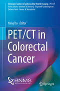
PET/CT in Colorectal Cancer
by Yong DuThis book is a pocket guide to the science and practice of PET/CT imaging of colorectal malignancies. The role of PET/CT in these patients, the characteristic PET/CT findings, and the advantages and limitations of this hybrid modality are all clearly described. In addition, information is provided on clinical presentation, diagnosis, staging, pathology, management, and radiological imaging. The book is published within the Springer series Clinicians' Guides to Radionuclide Hybrid Imaging, which is aimed at nuclear medicine/radiology residents and physicians, referring clinicians, radiographers/technologists, and nurses who routinely work in nuclear medicine and participate in multidisciplinary meetings. Compiled under the auspices of the British Nuclear Medicine Society, the series is the joint work of many colleagues and professionals worldwide who share a common vision and purpose in promoting and supporting nuclear medicine as an important imaging specialty for the diagnosis and management of oncological and non-oncological conditions.
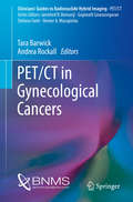
PET/CT in Gynecological Cancers
by Tara Barwick Andrea RockallThis book is a pocket guide to the science and practice of PET/CT imaging of gynecological malignancies. The scientific principles of PET/CT, the radiopharmaceuticals used in the context of gynecological cancers, the role of PET/CT in these patients, the characteristic PET/CT findings, and limitations and pitfalls are all clearly described. In addition, information is provided on clinical presentation, diagnosis, staging, pathology, management, and radiological imaging. The book is published within the Springer series Clinicians' Guides to Hybrid Imaging, which is aimed at referring clinicians, nuclear medicine/radiology physicians, radiographers/technologists, and nurses who routinely work in nuclear medicine and participate in multidisciplinary meetings. Compiled under the auspices of the British Nuclear Medicine Society, the series is the joint work of many colleagues and professionals worldwide who share a common vision and purpose in promoting and supporting nuclear medicine as an important imaging specialty for the diagnosis and management of oncological and non-oncological conditions.
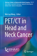
PET/CT in Head and Neck Cancer
by Wai Lup WongThis pocket book is an up-to-date guide to the diagnostic imaging of head and neck cancers. The focus is particularly on FDG PET/CT, with coverage of the basic principles, clinical indications, typical and atypical appearances, normal variations and artifacts, advantages, limitations, and pitfalls. Consideration is also given to emerging roles for PET/CT in head and neck cancer, including radiotherapy planning and treatment response monitoring, and to radiotracers beyond FDG. In addition, succinct information is provided on clinical presentation, diagnosis, staging, pathology, management, and other diagnostic imaging techniques. A brief discourse on the practice of guideline adoption is included. The book is published within the Springer series Clinicians' Guides to Radionuclide Hybrid Imaging (compiled under the auspices of the British Nuclear Medicine Society) and will be an excellent asset for clinicians, nuclear medicine physicians, radiologists, radiographers, technologists, and nurses who work in the field of head and neck cancer.
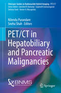
PET/CT in Hepatobiliary and Pancreatic Malignancies
by Nilendu Purandare Sneha ShahThis book is a pocket guide to the role of PET/CT in malignancies of the hepatobiliary system and pancreas. Imaging findings characteristic of malignancies are described and illustrated, and attention drawn to normal variants and artifacts. In addition, information is presented on epidemiology, clinical presentation, pathology, staging, and management in order to provide the clinical insight required by the imaging specialist. The book will be of immense value to practicing nuclear physicians as well as hybrid imaging specialists, particularly during reading sessions and multidisciplinary team meetings. It will also serve as a quick reference for residents and fellows in the PET/CT department. The book is published within the series Clinicians' Guides to Radionuclide Hybrid Imaging, which is compiled under the auspices of the British Nuclear Medicine Society. The series is the joint work of professionals worldwide who share a common vision in promoting nuclear medicine as an important imaging specialty for the diagnosis and management of oncological and non-oncological conditions.
PET/CT in Hodgkin’s Lymphoma
by Irfan KayaniThis book is a pocket guide to the practice of PET/CT imaging of Hodgkin's lymphoma. The role of PET/CT in Hodgkin's lymphoma, the characteristic findings and patterns, and the advantages and limitations of this hybrid modality are all clearly described. In addition, information is provided on clinical presentation, diagnosis, staging, pathology, management, and conventional radiological imaging. A useful pictorial atlas is included at the end of the book. PET/CT in Hodgkin's Lymphoma is published within the Springer series Clinicians' Guides to Radionuclide Hybrid Imaging, which is aimed at referring clinicians, nuclear medicine/radiology physicians, radiographers/technologists, and nurses who routinely work in nuclear medicine and participate in multidisciplinary meetings. Compiled under the auspices of the British Nuclear Medicine Society, the series is the joint work of many colleagues and professionals worldwide who share a common vision and purpose in promoting and supporting nuclear medicine as an important imaging specialty for the diagnosis and management of oncological and non-oncological conditions.
PET/CT in Infection and Inflammation (Clinicians’ Guides to Radionuclide Hybrid Imaging)
by Thomas Wagner Sandip BasuThis pocket book provides clinicians with the necessary information to understand the role of FDG PET/CT in infection and inflammation. It will help both in making appropriate imaging requests with adequate clinical information and in interpreting the report. The coverage encompasses a wide range of topics, including the role of PET/CT in pyrexia of unknown origin, vasculitis, autoimmune diseases, prosthetic joint infections, osteomyelitis and diabetic foot, immunodeficiency disease, and vascular graft surgery. The book will be a very useful guide to a great test that can provide significant assistance in patient management. It is published within the Springer series Clinicians’ Guides to Radionuclide Hybrid Imaging, in which leading professionals succinctly explain the importance of nuclear medicine in the diagnosis and management of oncological and non-oncological conditions.
PET/CT in Lung Cancer
by Archi Agrawal Venkatesh RangarajanThis concise, excellently illustrated pocket book provides an up-to-date summary of the science and practice of PET/CT imaging in lung cancer. The coverage encompasses the entire spectrum of lung cancer – pathology, radiological and PET/CT imaging, and management. Readers will also find information on the physics of PET and its use in respiratory gating and radiotherapy planning. The highlights of the book are the exquisite depiction of normal variants, pitfalls, and artifacts and a pictorial atlas of the various types of lung cancer and their manifestations. The contributing authors are well-known and experienced oncologists, pathologists, radiologists, and nuclear physicians. This book has been compiled under the auspices of the British Nuclear Medicine Society. It will be of high value for nuclear physicians, radiologists, referring clinicians and oncologists, and paramedical staff working in these fields
PET/CT in Lymphomas
by Athanasios D. Gouliamos John A. Andreou Paris A. KosmidisDrawing on an extensive series of cases, this book describes and illustrates in detail the FDG-PET/CT appearances of the most common lymphomas in both adults and children, covering presentations at various anatomic sites. In addition, all other aspects of the current application of FDG-PET/CT in the evaluation and management of patients with malignant lymphomas are described. Full explanations are provided of the potential benefits and limitations of advanced PET/CT techniques and technologies that support novel chemotherapy and radiotherapy approaches in the treatment of lymphoma, and particular attention is paid to the major challenge of incorporating progress in quantitative imaging technology into radiotherapy treatment planning, guidance, and monitoring. This clinical case-based atlas will be an invaluable tool for radiologists, hematologists, and clinical oncologists.
PET/CT in Melanoma
by Michael S. Hofman Rodney J. HicksThis pocket book provides up-to-date guidance on the use of PET/CT in patients with melanoma, which is of rapidly growing importance due to the emergence of immunotherapy. The role of PET/CT in diagnostic workup, staging, treatment selection, prognostication, and follow-up is clearly explained. Imaging features are described and illustrated with the aid of a series of teaching cases, and attention is drawn to normal variants, artifacts, and pitfalls. Readers will also find explanation of the relation of the clinical and pathological background to imaging and the value of PET/CT compared with conventional radiological imaging. The book is published within the Springer series Clinicians’ Guides to Radionuclide Hybrid Imaging (compiled under the auspices of the British Nuclear Medicine Society) and will be an excellent asset for referring clinicians, nuclear medicine/radiology physicians, radiographers/technologists, and nurses who routinely work in nuclear medicine and participate in multidisciplinary meetings.
PET/CT in Neuroendocrine Tumors
by Stefano Fanti Valentina AmbrosiniThis pocket book provides up-to-date descriptions of themost relevant features of neuroendocrine tumors (NETs) and the imagingmodalities currently available to assist specialists (clinicians, pathologists,radiologists, nuclear medicine physicians) in selecting optimal patientmanagement based on interdisciplinary collaboration. As the title indicates,the focus is particularly on PET/CT, with coverage of basic principles, theavailable radiopharmaceuticals, indications, typical and atypical appearances, normalvariants and artifacts, advantages, limitations, and pitfalls. In addition, succinctinformation is provided on the use of other imaging modalities, includingSPECT, CT, and MRI, and on pathology and treatment options. Imaging teachingcases are presented, and key points are highlighted throughout. The book ispublished as part of a series on hybrid imaging that isspecifically aimed at referring clinicians, nuclear medicine/radiology physicians,radiographers/technologists, and nurses who routinely work in nuclear medicineand participate in multidisciplinary meetings.
PET/CT in Non-Hodgkin Lymphoma (Clinicians’ Guides to Radionuclide Hybrid Imaging)
by Archi Agrawal Venkatesh Rangarajan Ameya D. PuranikThis book provides an up-to-date summary of the latest scientific developments on the use of PET-CT imaging in Non-Hodgkin Lymphoma (NHL). It encompasses the entire spectrum of NHL – from pathology to radiological and PET-CT imaging and to the management of NHL. The highlight of the book is the excellent pictorial depiction of normal variants, pitfalls and artifacts while exploring the different types of NHL and its manifestations. Each chapter, written by well-known and experienced oncologists and physicians, is enriched by a wide range of images to ensure a clear concept of PET-CT imaging in NHL and will definitely keep the readers interested.This book will be a useful tool for nuclear physicians, radiologists, referring clinicians and oncologists, as well as para-medical staff working in this field.
PET/CT in Oesophageal and Gastric Cancer
by Teresa A. SzyszkoThis book is a pocket guide to the science and practice of PET/CT imaging of esophageal and gastric malignancies. The scientific principles of PET/CT, the radiopharmaceuticals used in this context, the role of PET/CT, the characteristic PET/CT findings, and limitations and pitfalls are all clearly described. In addition, information is provided on epidemiology, clinical presentation, diagnosis, staging, pathology, management, and radiological imaging. The book is published within the Springer series Clinicians' Guides to Hybrid Imaging, which is aimed at referring clinicians, nuclear medicine/radiology physicians, radiographers/technologists, and nurses who routinely work in nuclear medicine and participate in multidisciplinary meetings. Compiled under the auspices of the British Nuclear Medicine Society, the series is the joint work of many colleagues and professionals worldwide who share a common vision and purpose in promoting and supporting nuclear medicine as an important imaging specialty for the diagnosis and management of oncological and non-oncological conditions.
PET/CT in Prostate Cancer
by Gary CookThis pocket book explains the significant and well-documented impact that PET/CT can have on the management of prostate cancer through the provision of high-quality evidence regarding function and structure. Up-to-date information is supplied on the relevance of PET/CT to diagnosis, treatment planning, and therapy, including the emerging role of PET/CT with PSMA. Readers will also find clear explanation of the relation of the clinical and pathological background to imaging and the value of PET/CT compared with conventional radiological imaging. The book will be an excellent asset for referring clinicians, nuclear medicine/radiology physicians, radiographers/technologists, and nurses who routinely work in nuclear medicine and participate in multidisciplinary meetings. It is published within the Springer series Clinicians' Guides to Radionuclide Hybrid Imaging, which presents contributions from professionals worldwide who share a common purpose in promoting nuclear medicine as an important imaging specialty for the diagnosis and management of oncological and non-oncological conditions.
