- Table View
- List View
Ultrasonic Topographical and Pathotopographical Anatomy: A Color Atlas
by O. V. Surnina Z. M. SeagalWritten by experienced and well-respected physicians and professors, this new all-color volume presents the ultrasonic topographical and pathotopographical anatomy of the body, including the head, neck, chest, anterolateral abdominal wall, abdominal organs, retroperitoneal space, male and female pelvises, and lower extremities. Specific and non-specific ultrasonic symptoms are suggested for normal and abnormal developmental variants, diffuse and local pathotopographical anatomy. This color atlas contains comparative topographical and pathotopographical data and is the first manual of its kind for students and medical specialists in different areas, including those specializing in medical sonography. The original technology was tested at clinics in patients subjected to ultrasonic monitoring. Because of early detection there were no false-positive or false-negative results. The therapy was effective, and, in some cases, the use of the original method of "seagalography" (optometry and pulsemotorgraphy) has made it possible to develop new methods of treatment and/or to determine the optimal doses of drugs, as well as to develop effective drug complexes for treatment of a given pathology. This important new volume will be valuable to physicians, junior physicians, medical residents, lecturers in medicine, and medical students alike, either as a textbook or as a reference. It is a must-have for any physician's library.
Ultrasonic and Electromagnetic NDE for Structure and Material Characterization: Engineering and Biomedical Applications
by Tribikram KunduMost books on nondestructive evaluation (NDE) focus either on the theoretical background or on advanced applications. Bridging the gap between the two, Ultrasonic and Electromagnetic NDE for Structure and Material Characterization: Engineering and Biomedical Applications brings together the principles, equations, and applications of ultrasonic and
Ultrasonics: Data, Equations and Their Practical Uses
by Dale Ensminger Foster B. StulenGain a Unique and Comprehensive Understanding of UltrasonicsDespite its importance, most books on ultrasonics cover only very specific sub-fields of the science. They generally also take a more mathematical approach and lack the wider scope needed to truly improve understanding and facilitate practical use of ultrasonics across a wide range of disc
Ultrasonographic Anatomy of the Face and Neck for Minimally Invasive Procedures: An Anatomic Guideline for Ultrasonographic-Guided Procedures
by Hee-Jin Kim Kwan-Hyun Youn Ji-Soo Kim You Soo Kim Sung Ok Hong Jongju NaThis is the very first book to describe the superficial anatomic structure of the face and neck by means of detailed ultrasonography (US). This superbly illustrated book will help aesthetic physicians to familiarize themselves with the US anatomy of the face and neck and to understand the applications and benefits of US when performing minimally invasive aesthetic procedures in this region. A deep understanding of anatomy is imperative if such procedures are to be safe and effective. Bearing in mind the range of potential anatomic variations, US can offer vital assistance in identifying target structures of the face beneath the skin when carrying out treatments that would otherwise be performed “blind”. In this book, readers will find detailed guidance on the use of US in the context of botulinum toxin and filler injections, threading procedures, and other minimally invasive aesthetic approaches. This is done with the aid of more than 530 US images, including cadaveric dissections and illustrations of volunteers and patients. For novices, valuable information is also provided on the basics of US imaging.
Ultrasonography Diagnosis of Peripheral Nerves: Cases and Illustrations
by Dingzhang Chen Minjuan ZhengAs a hot topic in ultrasound medicine, peripheral nerve ultrasound has its wide applications in clinical field. This book firstly introduces the anatomy of peripheral nerves, method and normal sonograms for peripheral nerve scanning. In the following chapters, common and typical cases of peripheral nerves diseases are presented with useful clinical information and relevant data, for example, ultrasound, MRI, clinical operation and pathology results. At the end of each disease, video with detailed explanation of diagnostic procedure and 2-3 bullet points in practical differential diagnosis are included to help readers taking notes. This book will be a valuable reference for physicians in ultrasound, anesthetists, neurologists, pain specialists, and practitioners interested in related field.
Ultrasonography for the Upper Limb Surgeon
by Thomas Apard Jean Louis BrasseurThis book combines orthopedists’ and radiologists’ perspectives to provide a comprehensive overview of the rapidly expanding use of ultrasound in orthopedic surgery. It also highlights the growing awareness of the potential of this non-invasive and portable, real-time imaging tool, which has led to its inclusion in the minimally invasive toolkit of upper limb surgeons.The book is divided into five parts – shoulder, elbow, forearm, hand and wrist and fingers. Each part focuses on a particular anatomic region or joint, carefully analyzing the sonoanatomy of its nerves, tendons and bones. For each region, experienced experts illustrate how to perform specific techniques under ultrasound control, ranging from classic procedures, like carpal tunnel release, to the treatment of less common conditions. Covering all the basic and practical aspects of this innovative, multi-disciplinary approach, as well as future perspectives, this unique book is a must-read for all orthopedists, radiologists, sports physicians and physiotherapist wanting to gain insights into this promising field.
Ultrasonography in Dentomaxillofacial Diagnostics
by Kaan OrhanThis book offers a comprehensive review of the state of the art in Ultrasonography (USG) dentomaxillofacial imaging to help radiologists and dentists in their training and daily practice.The book examines the relationship between clinical features, diagnosis, and choice of minimally invasive technique for a range of dentomaxillofacial disorders and provides information on post-treatment therapy. Accurate interpretation of indications for treatment is the cornerstone of success in medicine, and as such, the book explains how the selection of imaging technique is closely linked to clinical and diagnostic aspects and how recognition of this relationship forms the foundation for optimal outcomes. In addition to examining the various modalities, the book highlights the role of the latest USG imaging techniques. Further, it discusses in detail the pathology, treatment, and prognosis of common and rare diseases, as well as congenital/developmental malformations in the dentomaxillofacial, an area that is often underestimated and largely ignored by dentists.Featuring updated high-resolution images created with state-of-the-art equipment, the book introduces readers to current imaging modalities. It also includes pathological descriptions of radiologic diagnoses to help clarify the pathophysiology of the disease, while the pearls and pitfalls of image interpretation provide a quick reference guide for practitioners.Written by leading international experts, this outstanding book is a valuable resource for both radiologists, dentists and students seeking a more in-depth appreciation of the subject and its contribution to the scientific radiology community.

Ultrasonography in Gynecology
by FACS Botros R. M. B. Rizk Frcog Facog Hcld Frcs C Elizabeth E. PuscheckUltrasonography is a cornerstone in the evaluation of gynecologic disease. This authoritative new book looks at the techniques of ultrasonography in both office and hospital settings, offering guidance on the optimal use of equipment and covering the full range of benign and malignant gynecologic disease as well as infertility. Ultrasonography in Gynecology offers extensive coverage of the diagnostic potential of ultrasound in gynecologic disease, from the moment the patient walks into the physician's office. All the different approaches in the ultrasonographic evaluation of disease – including 3D ultrasonography, 3D sonohysterography, Doppler imaging and pelvic floor imaging – are extensively covered, with color images throughout. Written and edited by leaders in the field of ultrasonography who have actively participated in national and international teaching courses, Ultrasonography in Gynecology is a must for all gynecologists dealing with infertility, endometriosis, uterine fibroids, gynecologic cancers, and many more gynecologic conditions.
Ultrasonography in Reproductive Medicine and Infertility
by Botros R. M. B. RizkNowhere has the impact of ultrasonography been more dramatic than in reproductive medicine, particularly in the diagnosis of female and male infertility, the management of assisted reproductive procedures and the monitoring of early pregnancy. This authoritative textbook encompasses the complete role of ultrasonography in the evaluation of infertility and assisted reproduction. Covering every indication for ultrasonography in assisted reproductive technology, this will prove an invaluable resource in the evaluation of the infertile patient and optimization of the outcome of treatment. The interpretation of images to improve fertility and reproductive success is emphasized throughout. Ultrasonography in Reproductive Medicine and Infertility is essential reading for clinicians working both in IVF clinics and in office practice. It will be particularly useful to gynecologists, infertility specialists, ultrasonographers and radiologists working in reproductive endocrinology and infertility, assisted reproductive technology, ultrasonography and radiology.
Ultrasonography in Vascular Diagnosis
by Wilhelm Schäberle B. HerwigThis is the second edition of a well-received book that has been recommended for inclusion in any vascular library or vascular radiology suite. The first edition has been fully revised so as to provide a comprehensive, up-to-date account of vascular ultrasound that reflects recent advances. The emphasis remains on the clinical aspects most relevant to angiologists and vascular surgeons. Ultrasound anatomy is discussed, examination procedures explained, normal and pathological findings described, and the clinical impact of ultrasound assessed. Atlas sections present pertinent case material to illustrate typical ultrasound findings for both the more common vascular diseases and rarer conditions. This book will serve not only as an invaluable guide for beginners, but also as an indispensable reference for experienced sonographers, who will benefit from the detailed evaluation of the role of ultrasound as compared with other modalities and the discussion of ultrasound findings in their clinical context.
Ultrasonography in Vascular Diagnosis: A Therapy-oriented Textbook And Atlas
by Wilhelm Schäberle Bettina HerwigDer Band fasst die modernen Verfahren und neuesten Erkenntnisse auf dem Gebiet der Gefäßdiagnostik zusammen. Der Textteil beschreibt die Gefäßregion mit Sonoanatomie, Untersuchungsablauf und Normalbefund sowie die Indikation der Ultraschalluntersuchung und die klinische Relevanz der Untersuchungsergebnisse. Der Atlasteil veranschaulicht anhand aussagekräftiger Ultraschallbilder die verschiedenen Krankheitsbilder. Die 3. Auflage behandelt verstärkt die Bedeutung der Ultraschall-Routinediagnostik für seltene Gefäßerkrankungen.
Ultrasonography in the ICU
by Paula FerradaThis text presents a basic guide of the principles and applications of ultrasound in the critical care setting. The text also addresses the basic and clinical uses of ultrasound, including clinical cases at the end of each of the 7 sections of the clinical subdivisions. The final chapters focus on the issues of training, certification, credentialing and billing. These discussions make the text unique, as literature regarding monetary and training issues for ultrasound in the ICU is sparse. Each chapter is written by experts in the field, and supplemented with illustrations and links to videos of actual ultrasound examinations in patients. Ultrasonography in the ICU: Practical Applications will be of great utility to critical care specialists in the disciplines of surgery, medicine, anesthesiology and emergency medicine. In addition this book can be used as a teaching tool for critical care fellows and residents interested in further training in ultrasound for the critically ill patient.
Ultrasonography of the Hand in Rheumatology
by Peter Mandl Peter Vince BalintThis book is an essential guide for rheumatologists using ultrasound to study musculoskeletal structures and diagnose rheumatic diseases of the hand. A fundamental understanding of anatomy is key to identifying and interpreting pathological findings on imaging. This book provides comprehensive knowledge about both gross anatomy and, more importantly, comparative imaging anatomy, as depicted by ultrasound, X-ray, and MRI of the hand. This foundation enables clinicians to readily identify pathologic findings and thus improve diagnostic skills. The authors also offer step-by-step guidelines for using ultrasound, with comprehensive descriptions about body and transducer position, sonographic terminology, and anatomical landmarks in different planes of the hand. This book is an indispensable reference for rheumatologists and rheumatology residents using musculoskeletal ultrasound in clinical practice.
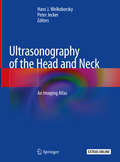
Ultrasonography of the Head and Neck: An Imaging Atlas
by Hans J. Welkoborsky Peter JeckerThis atlas presents a comprehensive and state-of-the-art overview of ultrasonography in the head and neck and will serve as a valuable resource for clinicians, surgeons, and otolaryngologists in private practice. The volume addresses all fields of office-based ultrasonography and gives an overview on the physical principles of ultrasound and sonographic techniques, along with detailed demonstrations of typical sonographic characteristics of particular diseases in the head and neck. Written by experts in the field it provides tips and tricks for ultrasound imaging. Subsequent chapters focus on office-based ultrasonography of the face and paranasal sinuses, salivary glands, floor of mouth and tonsil pathology, lymph node pathology, neck masses, thyroid and parathyroid glands, esophagus, and larynx. Special chapters address endosonography of the pharynx and larynx, interventional sonography, and intraoperative sonography. Latest technical developments in the field and their application to clinical ultrasonography are also demonstrated. A brief review of the existing latest literature addressing particular topics follow each chapter. All sonographic findings are demonstrated by high quality ultrasound-pictures and supplementary videos. Ultrasonography of the Head and Neck will serve as a useful guide for all physicians dealing with head and neck ultrasonography and its application to clinical medicine.
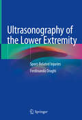
Ultrasonography of the Lower Extremity: Sport-Related Injuries
by Ferdinando DraghiThis book provides a detailed overview of ultrasound imaging of sport-related injuries of the lower extremity. The available literature focuses mainly on either clinical aspects or all imaging modalities and clinical aspects of sport-related pathologies, with little relevance on ultrasound. Indeed, recent advances in ultrasound technology, including high resolution, electronic, broadband transducers, have led to improved assessment of the musculoskeletal system, and ultrasound is now considered an optimal imaging technique to evaluate musculoskeletal sport-related injuries. Its advantages include the ability to perform dynamic examinations essential for many diagnoses, such as intrasheath instability of the peroneal tendons.Drawing on the author’s over 30 years of experience in clinical praxis, this book highlights the great potential of the ultrasonographic evaluation of sports-related injuries and is entirely devoted to this technique. Similar to the two previous monographs by the same author, the book has the form of an atlas-text, with a wealth of high-quality ultrasound images and schemes – a structure that has proved particularly effective for learning, especially for younger physicians. Ultrasonography of the lower extremity: sport-related injuries combines the interests of various specialists, including radiologists, physiatrists, orthopedists, rheumatologists, and ultrasound technicians.
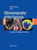
Ultrasonography of the Pancreas
by Mirko D'OnofrioUltrasonography (US) has long been considered an important diagnostic imaging modality for investigation of the pancreas despite certain significant and well-known limitations. Indeed, in many countries US represents the first step in the diagnostic algorithm for pancreatic pathologies. Recent years have witnessed major advances in conventional, harmonic, and Doppler imaging. New technologies, softwares, and techniques, such as volumetric imaging, enhancement quantification, and fusion imaging, are increasing the diagnostic capabilities of US. The injection of microbubble contrast agents allows better tissue characterization with definitive differentiation between solid and cystic lesions. Contrast-enhanced US improves the characterization of pancreatic tumors, assists in local and liver staging, and can offer savings in both time and money. Acoustic radiation force impulse (ARFI) imaging is a promising new US method to test, without manual compression, the mechanical strain properties of deep tissues. Furthermore, the applications and indications for interventional, endoscopic, and intraoperative US have undergone significant improvement and refinement. This book provides a complete overview of all these technological developments and their impact on the assessment of pancreatic pathologies. Percutaneous, endoscopic, and intraoperative US of the pancreas are discussed in detail, with precise description of findings and with informative imaging (CT and MRI) and pathologic correlations.
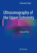
Ultrasonography of the Upper Extremity
by Ferdinando DraghiThis practice-oriented book, containing a wealth of high-quality ultrasound images, provides clear, concise, and complete coverage of the normal anatomy of the hand and wrist - tendons, nerves, and vascular structures - as well as the main pathologic conditions encountered in this area. The ultrasound images have been acquired with state of the art scanners and carefully labeled to facilitate recognition of each and every anatomic structure. Helpful comparison is also made with images and findings obtained using other diagnostic techniques, including in particular magnetic resonance imaging. The lucid text is complemented by practical tables summarizing key points for ease of reference. Readers will find Ultrasonography of the Upper Extremity to be a rich source of information on anatomy, examination techniques, and ultrasound appearances of one of the anatomic regions to have benefited the most from the technological revolution that has taken place in the field of ultrasonography during recent years. The book will appeal to both novice and experienced practitioners, including above all radiologists and ultrasound technicians but also rheumatologists and orthopedic surgeons. The author is the Director of the Operative Unit of Ecography at the Institute of Radiology, University Hospital Foundation IRCCS Policlinico San Matteo Pavia (Italy), and is Editor in Chief of The Journal of Ultrasound.
Ultrasonography of the Upper Extremity: Elbow
by Ferdinando DraghiThis book on elbow ultrasonography is a practice-oriented book, offering a wealth of high-quality ultrasound images, and providing clear, concise, and comprehensive coverage of the normal anatomy as well as the main pathologic conditions of the elbow. The ultrasound images have been obtained using state-of-the-art scanners and carefully labeled to facilitate recognition of each condition. The book also provides a helpful comparison of the images and findings obtained using other diagnostic techniques, including magnetic resonance imaging. The text is complemented by practical tables summarizing key points for ease of reference. Ultrasonography of the Upper Extremity: Elbow is a rich source of information on the anatomy, examination techniques and ultrasound appearances of one of the anatomic regions to have benefited most from the technological revolution that has taken place in the field of ultrasonography in recent years. The book appeals to both novice and experienced practitioners, including above all radiologists and ultrasound technicians, as well as rheumatologists and orthopedic surgeons.
Ultrasound Anatomy of Lower Limb Muscles
by Enzo Silvestri Davide Orlandi Alessandro MudaThe book provides a comprehensive description of the basic ultrasound principles, normal anatomy of the lower limb muscles and classification of muscle strain injuries. Ultrasound images are coupled with anatomical schemes explaining probe positioning and scanning technique for the various muscles of the thigh and leg. For each muscle, a brief explanation of normal anatomy is also provided, together with a list of tricks and tips and advice on how to perform the ultrasound scan in clinical practice. This book is an excellent practical teaching guide for beginners and a useful reference for more experienced sonographers.
Ultrasound Anatomy of Lower Limb Muscles: A Practical Guide
by Enzo Silvestri Davide Orlandi Alessandro MudaThe book provides a comprehensive description of the basic ultrasound principles, normal anatomy of the lower limb muscles and classification of muscle strain injuries. Ultrasound images are coupled with anatomical schemes explaining probe positioning and scanning technique for the various muscles of the thigh and leg. For each muscle, a brief explanation of normal anatomy is also provided, together with a list of tricks and tips and advice on how to perform the ultrasound scan in clinical practice. This book is an excellent practical teaching guide for beginners and a useful reference for more experienced sonographers.
Ultrasound Assessment in Gynecologic Oncology
by Juan Luis AlcázarThis innovative guide will help gynecologists or gynecologic surgeons to monitor the staging and progress of oncology treatment. An international expert here shows what office ultrasound can be used to achieve, how it correlates with other clinical findings, and how it can be integrated as necessary with other modalities and with the latest technological advances and developments in the field.
Ultrasound Diagnostics of Thyroid Diseases
by Alexander N. Sencha Yury K. Alexandrov Yury N. Patrunov Denis V. Belyaev Mikhail S. Mogutov Peter M. Kotlyarov Vladimir P. KharchenkoAs the basis for this book, the authors have analyzed more than 100,000 ultrasound examinations performed between 1995 and 2008 in patients with thyroid and parathyroid disease, as well as many thousands of diagnostic and therapeutic ultrasound-guided minimally invasive procedures. The opening chapters include discussion of current ultrasound techniques, pitfalls, and ultrasound examination of the thyroid in children. Detailed attention is then devoted to findings in the normal thyroid and in the presence of diffuse and focal changes. Further chapters focus on such topics as the role of ultrasound after thyroid surgery and ultrasound diagnosis of parathyroid disease, recurrent goiter, and neck masses. Ultrasound-guided minimally invasive techniques, such as fine-needle aspiration biopsy and percutaneous laser ablation, are considered in depth. This up-to-date and richly illustrated book will interest and assist specialists in ultrasound diagnostics, radiologists, endocrinologists, and neck surgeons.
Ultrasound Energy and Data Transfer for Medical Implants (Analog Circuits and Signal Processing)
by Catherine Dehollain Francesco MazzilliThis book presents new systems and circuits for implantable biomedical applications, using a non-conventional way to transmit energy and data via ultrasound. The authors discuses the main constrains (e.g. implant size, battery recharge time, data rate, accuracy of the acoustic models) from the definition of the ultrasound system specification to the in-vitro validation.The system described meets the safety requirements for ultrasound exposure limits in diagnostic ultrasound applications, according to FDA regulations. Readers will see how the novel design of power management architecture will meet the constraints set by FDA regulations for maximum energy exposure in the human body. Coverage also includes the choice of the acoustic transducer, driven by optimum positioning and size of the implanted medical device. Throughout the book, links between physics, electronics and medical aspects are covered to give a complete view of the ultrasound system described.Provides a complete, system-level perspective on the use of ultrasound as energy source for medical implants;Discusses system design concerns regarding wireless power transmission and wireless data communication, particularly for a system in which both are performed on the same channel/frequency;Describes an experimental study on implantable battery powered biomedical systems;Presents a fully-integrated, implantable system and hermetically sealed packaging.
Ultrasound Fundamentals: An Evidence-Based Guide for Medical Practitioners
by Nalini Vadivelu Alan David Kaye Jinlei Li Robert Ming-Der ChowWritten by experts in the field, this concise and evidence-based ultrasound text includes key topics ranging from the head and neck to the upper and lower extremity, covering all the clinically relevant sonoanatomy. This 33-chapter book emphasizes the practical use of ultrasound for the diagnosis and treatment of a multitude of conditions in various specialty areas such as airway management, cardiovascular disease assessment, pulmonary status evaluation, orthopedics, gynecology and pediatrics. The optimal techniques and the step-by-step interpretation of normal and pathologic sonoanatomy are discussed in detail. This text can be used as a starting point for the study of ultrasound guided diagnosis and treatment, a refresher manual for sonoanatomy on major organ systems, or a last-minute guide before a bedside procedure. There is a great breadth of material that is covered in a comprehensive manner, making it a great resource for board review and exam preparation for various medical, surgical and allied specialties. Unique and pragmatic, Ultrasound Fundamentals is a back to basics manual on normal and pathologic sonoanatomy of head and neck, upper and lower extremity, chest, abdomen and other major organ systems
Ultrasound Guided Vascular Access: Practical Solutions to Bedside Clinical Challenges
by Matthew D. Ostroff Mark W. ConnollyThis casebook covers ultrasound guided vascular access with a focus on patient safety. Vascular access is the catalyst between provider and patient. Initially, patients were treated at hospitals for grave illnesses or terrible accidents; however, due to significant strides in medicine, the emerging hospitalized patient is now more commonly treated for chronic illnesses. For these patients, repeated hospital visits slowly deplete their vasculature, creating one of modern medicine’s greatest obstacles: vascular access. This book provides safe solutions to bedside clinical challenges from peripheral to tunneled central lines in the neonatal, pediatric, and adult patient populations.Chapters present unique patient cases, incorporating the latest technology and techniques for safely and methodically meeting vascular access challenges, detailed discussions on therapeutic interventions, and trouble shooting. The book is structured so that each chapter builds on the last, offering peripheral, central, and tunneled catheter solutions through a standardized approach. Finally, each part features a COVID-19 case study including vascular access performed in the prone position. This is an ideal guide for trainees, students, clinicians, and healthcare professionals performing ultrasound guided vascular access on patients.