- Table View
- List View
Ultrasound Imaging
by Jasjit S. Suri Andrew F. Laine João Miguel SanchesDiagnostic and Therapeutic Ultrasound has recently taken an explosive growth for better safer, economic, mobile and high quality healthcare. This technology is very appealing for medical applications because it is non-ionizing, non-invasive and it is available in most of the medical and clinical facilities. Its low cost, when compared with other medical image modalities, makes it one of the preferred tools for medical monitoring, follow-up and diagnosis. Besides the traditional fields of Cardiology and Obstetrics, where it is extensively used for long time, it has became also very useful in the diagnosis of diseases of the prostate, liver and coronaries and carotids atherosclerosis. However, Ultrasound images present poor quality, very low signal to noise ratio and a lot of artifacts. The extraction of useful information from Ultrasound data for diagnosis is a challenge task that makes this medical image modality a very active field of research. The difficulties are being overcome and novel and advanced methods are being proposed for detection, characterization and segmentation of abnormalities in several organs. In fact, Ultrasound application range is vast, covering almost all organs of the human body, including the brain where Tran-cranial Doppler Ultrasound is very important to assess the brain vasculature. This book presents some of the recent advances in Ultrasound imaging technology covering several organs and techniques in a Biomedical Engineering (BME) perspective. The focus of the book is in the algorithms, methodologies and systems developed by multidisciplinary research teams of engineers and physicians for Computer-Aided Diagnosis (CAD) purposes. Cardiovascular and Cancer, the most common life-threatening diseases in western countries, are two of the most important topics focused in the book. However, other advanced issues are also presented such as Intravascular Ultrasound, 3D US and Ultrasound in Computer-Aided Surgery (CAS). Some chapters are direct contributions from medical research groups where Ultrasound has also received great attention in the last decade. By this, new techniques based on Ultrasound were introduced in the clinical practice for diagnosis and therapeutics, mainly in hospital facilities.
Ultrasound Imaging and Therapy (Imaging In Medical Diagnosis And Therapy Ser.)
by Aaron Fenster James C. LacefieldUp-to-Date Details on Using Ultrasound Imaging to Help Diagnose Various DiseasesDue to improvements in image quality and the reduced cost of advanced features, ultrasound imaging is playing a greater role in the diagnosis and image-guided intervention of a wide range of diseases. Ultrasound Imaging and Therapy highlights the latest advances in usin
Ultrasound Imaging in Reproductive Medicine
by Laurel Stadtmauer Ilan Tur-KaspaOver the last 25 years, the advances in ultrasound have paralleled advances in Assisted Reproductive Technology (ART). ART could not even be practiced or considered today without imaging. Ultrasound has become the most important and widely used tool in the diagnosis and treatment of infertility. Ultrasound evaluation is one of the first steps to assess the cause of infertility; the three areas of evaluation are the ovaries, uterus, and fallopian tubes. Ultrasound allows physicians to diagnose ovarian reserve but also pathologies such as polycystic ovarian syndrome, endometriosis, or other ovarian cysts that can impact fertility. The results of this initial exam immediately affect the decisions in the management of the patient's condition. When fertility treatments begin, ultrasound is used in almost any interaction with the patient in order to monitor follicular development and endometrial response; ultrasound guidance is also vital for embryo retrieval and transfer (ET). Ultrasound Imaging in Reproductive Medicine provides a comprehensive survey of the use of ultrasonography in the female pelvis for physicians, nurses, and ultrasonographers actively involved in reproductive medicine and infertility. With a critical evaluation of advantages and disadvantages, the book covers traditional and new technologies, including three-dimensional (3D) ultrasound for ovarian reserve, ovarian monitoring and endometrial cavity assessment, Ultrasound, MRI and CT evaluation of tubal patency, MRI guided ultrasound procedures for treatment of uterine fibroids, imaging techniques of the embryo and embryo transfer, and pulsed color Doppler techniques. 3D ultrasound to assess ovarian and endometrial volume and 3D automated monitoring of follicles are also covered in detail with up to date references.
Ultrasound Imaging in Reproductive Medicine: Advances in Infertility Work-up, Treatment and ART
by Ilan Tur-Kaspa Laurel A. StadtmauerNow in an updated edition, this is the most comprehensive book on modern ultrasound imaging in assisted reproductive technology (ART) and reproductive medicine. Fully revised and expanded, it covers emerging technologies possible with the improvement in ultrasound equipment. 3-D monitoring of ovarian follicles, bidirectional vibrant color and Doppler, and improved 3-D and 4-D imaging of reproductive structures are discussed. MRI-guided ultrasound procedures are covered, and comparisons of 3-D imaging with MRI imaging for uterine anomalies is reviewed with an emphasis on the advantages of 3-D performed in the gynecologist’s office, and as a less expensive modality.The overall approach of the original edition is maintained, starting with ultrasound safety and technique and diagnosis of the ovary, uterus and fallopian tubes (both normal and pathologic), followed by both male and female infertility and ART treatments and procedures. Ultrasound monitoring of follicular development, the endometrium, and as an aid in embryo transfer to maximize IVF success rates are reviewed. Topics new to this edition include updated information on the diagnosis of benign and malignant adnexal masses, 3-D follicle monitoring, and the diagnosis of adenomyosis and endometriosis, including deep inseminated endometriosis. Additionally, the evaluation of endometrial receptivity, the use of contrasts for fallopian tube patency, controversies regarding septate uterus versus arcurate uterus with the use of 3-D ultrasound, and 3-D ultrasound with saline infusion sonogram and early pregnancy ultrasound are all discussed.An excellent resource for reproductive medicine and ART specialists, gynecologists and ultrasonographers alike, Ultrasound Imaging in Reproductive Medicine, Second Edition covers all that clinicians need to know about the role of ultrasound, from the first time a woman comes into the clinic for treatment, including ART, to early pregnancy monitoring. See better, do ART better.
Ultrasound Protocol for Facial Aesthetics: A practical clinical guide
by Kathryn Malherbe Stefania RobertsThis book provides a systematic approach to facial ultrasound, using anatomical landmarks and targeting common facial muscles during aesthetic treatment. It also includes the most common clinical indications and complications found during aesthetic therapy. The various muscles have been grouped into the upper, mid and lower face and are also classified as either easy, intermediate or advanced in the level of experience a user requires to discern these muscles on ultrasound. High-frequency ultrasound in the field of radiology has progressed to other fields of medical treatment, most notably within aesthetic medicine in recent years. Facial ultrasound has been limited to the clinical assessment of varying skin conditions, as the diagnosis and treatment have been primarily based on dermatological assessment. Having said that, the recent increase in aesthetic treatment methods has led to a rising number of complications, risks, and poor outcomes. The singular use of surface anatomy and clinical presentation of a patient before fillers are used has proven to be limited in assessing the risks prior to treatment. For this reason, ultrasound of the face has allowed clinicians to develop a keen sense of vascular presence, asymmetry of muscles, and also to help determine the depth for needle-guided injections.
Ultrasound Scoring of Joint Synovitis: Objective Quantification of Articular Inflammation
by Ami Ben-Artzi Gurjit S. Kaeley Veena K. RanganathA common manifestation of many autoimmune diseases is joint inflammation. The degree of inflammation of the joints is often used as a metric of disease activity. Assessing disease activity has diagnostic, therapeutic, and prognostic implications. Inflammation of the joints is often used as a proxy for inflammation in other tissue, and assessing this inflammation has implications for the health of numerous organs in in the body. Currently, physicians use palpation of joints to assess for joint inflammation. This method, while accessible and easy to apply, is lacking in precision. Ultrasound has also been used to assess for joint inflammation for over 20 years, but applying ultrasound technology has proved challenging. There is lack of agreement on optimal joint view, which joints should be examined in which disease, and how inflammation should be graded. Various scanning protocols and grading schemes have been proposed, but each has its strengths and weaknesses. Ultrasound Scoring of Joint Synovitis is based off the work of three authors who tackled the limitations of contemporary inflammation scoring systems, and developed an ultrasound-based inflammation scoring system which can be applied with consistency and reproducibility. In their research, the authors scanned thousands of joints and spent thousands of hours discussing the scans. They produced a scoring system which will allow the user of the atlas to grade joint inflammation in a way that is less subjective and more reproducible than the current systems used. Objective descriptions allow the user to quantify sonographic signs of joint inflammation with an intentional bias for the more clinically relevant features of ultrasound pathology. For students of musculoskeletal ultrasound, the book offers detailed descriptions of the sonographic appearance of every level of joint inflammation. For researchers, the book, and the system laid out in the book, are truly state-of-the-art and will enable better precision in research and clinical care of patients with joint inflammation.
Ultrasound Technology for Clinical Practitioners
by Crispian OatesUltrasound Technology for Clinical Practitioners A hands-on and practical roadmap to ultrasound technology for clinical practitioners who use it every day In Ultrasound Technology for Clinical Practitioners, distinguished medical physicist and vascular ultrasound scientist Crispian Oates delivers an accessible and practical resource written for the everyday clinical user of ultrasound. The book offers complete descriptions of the latest techniques in ultrasound, including ultrafast ultrasound and elastography, providing an up-to-date and relevant resource for educators, students, and practitioners alike. Ultrasound Technology for Clinical Practitioners uses a first-person perspective that walks readers through a relevant and memorable story containing necessary information, simplifying retention and learning. It makes extensive use of bulleted lists, diagrams, and images, and relies on mathematics and equations only where necessary to illustrate the relationship between other factors. Physics examples come from commonly known contexts that readers can relate to their everyday lives, and additional description boxes offer optional, helpful info in some topic areas. Readers will also find: A thorough introduction to the foundational physics of ultrasound, as well as the propagation of the ultrasound pulse through tissue Comprehensive discussions of beam shapes, transducers, imaging techniques, and pulse echo instrumentation In-depth examination of image quality and artefacts and the principles of Doppler and colour Doppler ultrasound Fulsome treatments of measurement taking and safety and quality assurance in ultrasound Perfect for sonographers, echocardiographers, and vascular scientists, Ultrasound Technology for Clinical Practitioners will also earn a place in the libraries of radiologists, cardiologists, emergency medicine specialists, and all other clinical users of ultrasound.
Ultrasound and Carotid Bifurcation Atherosclerosis
by Efthyvoulos Kyriacou Kirk W. Beach Constantinos S. Pattichis Andrew NicolaidesUltrasound and Carotid Bifurcation Atherosclerosis provides a comprehensive overview of the most recent advancements in instrumentation, imaging techniques including the use of contrast enhancement agents, plaque image analysis and its automation, elastography and plaque motion analysis; also, the use of ultrasonic and other biomarkers in the detection of the high risk cardiovascular individual. Finally, it deals with the application of IVUS, TCD and carotid plaque characterization in clinical practice and in stroke risk stratification. Ultrasound and Carotid Bifurcation Atherosclerosis is intended for all those working in the field of atherosclerosis, ultrasound imaging and cardiovascular risk, including the clinician, the vascular ultrasonographer, the epidemiologist, the molecular biologist, the biomedical engineer and the informatics scientist. Furthermore, this book bridges the gap between the researcher and the clinician, who is keen to incorporate the latest results of research to his daily practice.
Ultrasound and Infertility
by Asim KurjakA comprehensive survey of the use of ultrasound in management of infertile patients is presented in this publication. Particular atten-tion is given to recently developed techniques such as assessment of endometrial changes, ovarian blood flow measurements, and per-cutaneous oocyte retrieval for in vitro fertilization. The very re-cent technique of transvaginal sonography is presented and richly illus-trated with original results obtained in biopsy-guided oocyte re-trieval, and in the precise delineation of follicle size and number for infertility treatment. Guidance in the interpretation of ultrasonic findings, which include potential limitations and pitfalls, is provided in each chapter. Researchers and practitioners interested in the management of infertile patients will find this volume indispensable.
Ultrasound for Interventional Pain Management: An Illustrated Procedural Guide
by Philip Peng Roderick Finlayson Sang Hoon Lee Anuj BhatiaDue to a wide-spread developing interest in ultrasound-guided pain intervention by clinicians, the demand for a practical reference material on this topic has grown simultaneously. This book thoroughly satisfies the need for such a reference, as it contains text written by experts in the field and a multitude of unique, educational illustrations. Spinal pain, the musculoskeletal system, and peripheral structures function as the fundamental items of discussion across three divided sections. In order to augment the reader’s learning experience, the high-quality images found within each chapter provide step-by-step guidance on the various ultrasound scanning procedural processes. Additionally, tips and pearls for scan and injection supplement each chapter conclusion. Ultrasound for Interventional Pain Management: An Illustrated Procedural Guide is a pragmatic, indispensable resource that helps interested clinical practitioners enhance their visual memory and overall understanding of this method.
Ultrasound for Primary Care (M - Medicine Ser.)
by Paul BornemannMaster high-yield point-of-care ultrasound applications that are targeted specifically to answer questions that arise commonly in the outpatient clinic! Written for primary care providers in Family Medicine, Pediatrics and Internal Medicine, Ultrasound for Primary Care is a practical, easy-to-read guide. Learn to incorporate ultrasound to augment your physical exam for evaluation of thyroid nodules, enlarged lymph nodes, pericardial effusion, chronic kidney disease, and a host of musculoskeletal issues, and much more. Additionally, included are chapters on ultrasound for guidance of procedures including joint injections, lumbar puncture and needle biopsy, to name a few. Well-illustrated and highly templated, this unique title helps you expand the scope of your practice and provide more effective patient care.
Ultrasound in Assisted Reproduction and Early Pregnancy
by Arianna D’Angelo and Nazar N. AmsoThis text offers a succinct overview of the essential clinical applications of ultrasound in infertility management. It will be of benefit to established practitioners in reproductive medicine, as it details the aspects of quality, safety, training, and certification that help improve standards of practice. Those in training or with a special interest in fertility issues will also find it essential reading. Print versions of this book also include access to the eBook version with links to procedural videos.
Ultrasound in Assisted Reproduction and Early Pregnancy: A Practical Guide
by Jane S. Fonda Rachael J. Rodgers William L. LedgerUltrasonography is a crucial tool in successful assisted reproduction but requires a steady hand and can often be difficult for unconfident clinicians. A comprehensive ultrasound imaging reference, this is an essential guide for trainee clinicians, ultrasonographers, and nurses working in the field of assisted reproductive technology. Providing the reader with an overview of the process and a foundation to direct their ultrasound assessment of each patient, it contains highly practical tips and tricks for obtaining the best images. Heavily illustrated with example images, the role of ultrasound in fertility treatment is explained, as well as how to identify the uterus and ovaries, measure the endometrium, count follicles and recognize pathology. The role of ultrasound in assisted reproduction is covered, including transvaginal oocyte collection, embryo transfer, early pregnancy, miscarriage and ectopic pregnancy. This is an indispensable reference for clinicians new to ultrasound in assisted reproduction.
Ultrasound in Gynecology
by Mala SibalThis atlas and guide book is focused on gynecological ultrasound, an area that has remained in the shadow of obstetric ultrasound & fetal medicine. Gynecological ultrasound has seen rapid advances owing to expanding research and improved ultrasound equipment. This book leverages these advances and provides abundant illustrations and practice points of classical and new ultrasound features. It serves as a guide for radiologists, gynecologists and sonologists for the accurate diagnosis of gynecological pathologies. The chapters of this book also serve as a comprehensive resource for various topics with hundreds of images and figures, including basic gray scale images, Doppler studies and three dimensional ultrasound illustrations. In addition, standard terms for the evaluation and reporting of gynecological pathologies are discussed. Emergencies like ovarian torsion, complex adnexal cyst are also covered.
Ultrasound in Peripheral, Neuraxial and Perineuraxial Regional Anaesthesia
by Eryk Eisenberg Elisabeth GaertnerThis comprehensive, highly didactic book on ultrasound-guided regional anesthesia (peripheral, neuraxial and perineuraxial nerve blocks) presents meticulously labelled images, diagrams and picture-in-picture samples and includes high-quality, vignetted illustrations that are consistent in style. The ultrasound images are outstanding and carefully selected to demonstrate the most clinically relevant situations. Importantly, they have a real-world appearance, including actual needle paths and desired disposition of injectate during nerve block procedures; most are from the original database of Dr. Eisenberg. All the supplementary material is authoritative and presented as an artful balance of years of clinical experience and a summary of the peer reviewed literature.Ultrasound in Peripheral, Neuraxial and Perineuraxial Regional Anaesthesia, accompanied by richly illustrated material and videos of state-of-the-art techniques, is of interest to anyone interested in learning, furthering their existing knowledge of, or teaching ultrasound-guided regional anesthesia.
Ultrasound in Reproductive Healthcare Practice
by Paula Briggs Mary Pillai Julie-Michelle BridsonChallenge your knowledge of ultrasound to address sexual health abnormalities and early pregnancy issues, alongside identifying, classifying and managing a wide range of gynaecological conditions, with this essential manual. Authored by experts in reproductive health, this bespoke guide delivers practitioners of all levels with a broad scope of sexual and reproductive disorders, as captured by ultrasound. Presenting operational issues and suggested training, this textbook ensures high-quality care in gynaecology, sexual and reproductive health and pregnancy advisory services. For use in a traditional hospital setting through to more remote locations, this guide provides an invaluable toolkit for trainees, sonographers, nurses and clinicians worldwide. Offering clear clinical ultrasound images and extensive case studies with a focus on pregnancy advisory services, this adaptable textbook provides reliable support for those who are in contact with common, rare and understudied reproductive conditions, wishing to achieve successful diagnosis and optimal imaging first time. Bespoke and wide-ranging, this guide encompasses a range of understudied medical gynaecological conditions and early pregnancy issues, through to contraception and sexually transmitted infections. The first of its kind in using ultrasound as a tool to address conditions specific to sexual reproductive healthcare. Formalises ultrasound practice within sexual reproductive healthcare for the hospital setting, and remote settings, away from mainstream radiology services. Includes clear illustrations of clinical and ultrasound images, and case studies tailored to challenging pregnancy advisory services.
Ultrasound in Rheumatology: A Practical Guide for Diagnosis
by Qasim Akram Subhasis BasuThis book provides a practically applicable manual to the utilisation of ultrasound in rheumatology. Each chapter includes high-quality diagrams of each anatomical region covered, accompanied by an ideal scan with written and pictorial demonstrations, as well as an ideal ultrasound image, that has been obtained via a high-end machine for optimal image quality. This systematic approach to describing the application of ultrasound in rheumatology enables the reader to develop a deep understanding of how to correctly make use of ultrasound technologies in their daily practice.Ultrasound in Rheumatology: A Practical Guide for Diagnosis is an easy to follow guide to the application of ultrasound in rheumatology and is a valuable resource for the trainee and practising rheumatologist seeking a guide on the correct use of ultrasound.
Ultrasound in the Critically Ill: A Practical Guide
by Andrew Campbell Ashley Miller Andrew Walden Matthew WiseThis book provides a practically applicable guide to the use of ultrasound in the care of acutely and critically ill patients. It is laid out in two sections. The first section attempts to take a comprehensive approach to specific systems of examination taking an organ focused approach covering techniques including Focussed Assessment with Sonography for Trauma (FAST) scanning and venous sonography. The second section presents a range of specific cases enabling the reader to develop an understanding of how to apply these methodologies effectively into their day-to-day clinical practice.Ultrasound in the Critically Ill: A Practical Guide describes how to use ultrasound technologies in day-to-day clinical practice. Therefore, it is an ideal resource for all trainee and practicing physicians who utilize these technologies on a day-to-day basis.
Ultrasound of Congenital Fetal Anomalies: Differential Diagnosis and Prognostic Indicators
by Dario Paladini Paolo VolpeAn acclaimed overview of ultrasound for the prenatal diagnosis of congenital anomalies returns in a new enlarged edition. In particular, the coverage of both Central Nervous System congenital and acquired anomalies as well as Congenital Heart Disease has been expanded enormously, to make this an impressive comprehensive resource for Fetal Neurology and Fetal Cardiology. Together with additional new chapters on guidelines and protocols, equipment, and disorders of sexual differentiation, and new insight into fetal surgery procedures, this third edition almost becomes three books in one.
Ultrasound of Congenital Fetal Anomalies: Differential Diagnosis and Prognostic Indicators, Second Edition
by Dario Paladini Paolo VolpeThe most frequently asked questions that confront the fetal medicine trainee/expert on a daily basis are “Is the finding real or merely an artifact?” and “Is the diagnosis correct?”. However, to be able to find the description of an abnormal ultrasound finding in a textbook, one generally has to search by the definite diagnosis, which has not been done as yet. This uneasy feeling was the first factor that directed the layout of Ultrasound of Congenital Fetal Anomalies: Differential Diagnosis and Prognostic Indicators, Second Edition. Copiously illustrated, the book displays fetal anomalies by scanning view and descriptions of all major ultrasound planes, detailing what can be considered a normal view and what cannot. See What’s New in the Second Edition: Early detection of fetal anomalies (1214 weeks) Ultrasound in fetal infections and in twins The nuchal translucency issue, the newest intracranial translucency as well as the range of congenital anomalies detectable at this gestational age Expanded coverage of heart anomalies, including arrhythmias and early fetal echocardiography The author’s mission continues to be to provide guidance on how to quickly recognize and diagnose congenital fetal anomalies, beginning at the beginning with ultrasound sigh all the way through to final diagnosis.
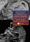
Ultrasound of Mouse Fetal Development and Human Correlates (Reproductive Medicine and Assisted Reproductive Techniques Series)
by Mary C. Peavey Sarah K. Dotters-KatzFetal development in the mouse is routinely and increasingly utilized for advancing translational research and medical innovation for human obstetrical care. This is the first and only manual to provide necessary content on how this should be handled for accurate and effective data collection. Detailed descriptions and examples demonstrate how researchers and clinicians can use murine fetal and obstetrical data to improve future human applications in diseases such as infertility, recurrent pregnancy loss, intrauterine fetal growth restriction, placental insufficiency, and intrauterine fetal demise, as well as organ-specific developmental disease.
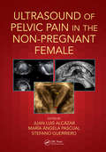
Ultrasound of Pelvic Pain in the Non-Pregnant Patient
by Stefano Guerriero Juan Luis Alcázar María Ángela PascualThere are many possible causes of pelvic pain in a non-pregnant female patient, and it has been estimated to be responsible for nearly 40% of all visits by female patients to a family doctor and 10% of all referrals to specialist gynecologists. However, the topic of how to investigate and diagnose has been surprisingly neglected in print. This important and much-needed text from an internationally respected expert shows how important ultrasound can be as a tool for physicians caring for women's health.
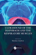
Ultrasound of the Diaphragm and the Respiratory Muscles
by Massimo ZambonUltrasound is the most reliable, easily available, fast, non-invasive technique to study diaphragm function, and is an irreplaceable tool to diagnose, monitor, and follow -up critical respiratory patients. This essential guide analyses every aspect of ultrasound of the diaphragm and respiratory muscles, a reliable assessment whose function is vital to delivering the most suitable treatment. Ultrasound of the Diaphragm and the Respiratory Muscles also provides insight to diagnosing diaphragmatic dysfunction or paralysis following surgery or neuromuscular diseases, to follow the muscular activity and the time-course of atrophy during mechanical ventilation, and to monitor the weaning phase. It is ideal for professionals and trainees practiscing ultrasound in a clinical setting. Key Features Sets the standard for training and competency of this emerging, yet scientifically approved non-invasive technique of ultrasound with all the essential information on how to perform ultrasound and interpret the images obtained. Features clear and didactic images demonstrating echo findings in various situations along with videos of diaphragmatic ultrasound offering a unique "window" on mechanically ventilated patients, allowing to take important clinical decisions on ventilatory modes and assistance by pulmonologists, critical care specialists, thoracic surgeons, emergency medicine specialists, as well as trainees. Includes a chapter on paediatric ultrasound along with ultrasound of other respiratory muscles (i.e., intercostal and abdominal) which is emerging as a useful complementary tool.
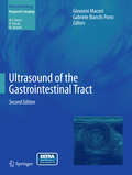
Ultrasound of the Gastrointestinal Tract
by Giovanni Maconi Gabriele Bianchi PorroThis is the second, updated and extended edition of a well-received book that offers a comprehensive overview of ultrasonographic imaging of acute and chronic gastrointestinal diseases, including acute abdomen, appendicitis, diverticulitis, inflammatory bowel diseases, neoplasms and masses, infections, malabsorption syndromes, and rare conditions. The value of ultrasound in each disorder is clearly explained and illustrated, and limitations identified. Information is also provided on recent technical developments and ultrasound applications that are likely to become of increasing importance, such as functional and 3D ultrasound, contrast agents and intraoperative ultrasound, elastography, and transperineal ultrasound. The authors are all distinguished experts in the topics they address. Ultrasound of the Gastrointestinal Tract will be a helpful guide in daily practice not only for radiologists but also for gastroenterologists, abdominal surgeons, pediatricians, and oncologists.
Ultrasound of the Testis for the Andrologist: Morphological And Functional Atlas (Trends In Andrology And Sexual Medicine Ser.)
by Andrea Lenzi Andrea M. IsidoriThis book presents a comprehensive study of scrotal ultrasound, helping readers cope with the growing number of pathology pictures revealed by accurate ultrasound examinations, and highlighting the novel applications of contrast-enhanced ultrasonography and elastography.This unique reference guide to scrotal ultrasonography draws on the accumulated expertise of the Experimental Medicine Department at “Sapienza” University, where the andrological ultrasonography unit has performed over 10,000 testicular ultrasound examinations for various conditions and explored experimental new imaging techniques. This core experience has been enriched by insightful contributions from several international experts to form one of the most comprehensive collections of ultrasound images, many in full color, of scrotal pathology in the world.The book’s emphasis on functional interpretation of the images, supplemented by clinical data, make it a unique tool for clinical management. This approach is intended to increasingly familiarize clinicians with the potentials of ultrasonography, from the basics to the most advanced approaches, so as to encourage them to incorporate this examination as a central component of the diagnostic pathway