- Table View
- List View

Anatomy and Physiology
by Eddie Johnson J. Gordon Betts Kelly A. Young Peter Desaix Jody E. Johnson Oksana Korol Dean Kruse Brandon Poe James A. Wise Mark WombleAnatomy and Physiology 2e is developed to meet the scope and sequence for a two-semester human anatomy and physiology course for life science and allied health majors. The book is organized by body systems. The revision focuses on inclusive and equitable instruction and includes new student support. Illustrations have been extensively revised to be clearer and more inclusive. The web-based version of Anatomy and Physiology 2e also features links to surgical videos, histology, and interactive diagrams. Please learn more about the changes by previewing the preface.
Anatomy and Physiology
by Frederic H. Martini Judi L. NathThis new textbook answers the need for a briefer version of Martini's Fundamentals of Anatomy & Physiology for the 2-semester anatomy and physiology course. With condensed explanations and less detailed discussions, this slim volume retains Martini's award-winning art program, key clinical discussions, and clear, straightforward writing style. Accompanied by a first-rate, text-specific supplements package, Anatomy & Physiology provides instructors and students with a compact and efficient learning system.

Anatomy and Physiology
by OpenStaxAnatomy and Physiology 2e is developed to meet the scope and sequence for a two-semester human anatomy and physiology course for life science and allied health majors. The book is organized by body systems. The revision focuses on inclusive and equitable instruction and includes new student support. Illustrations have been extensively revised to be clearer and more inclusive. The web-based version of Anatomy and Physiology 2e also features links to surgical videos, histology, and interactive diagrams. Please learn more about the changes by previewing the preface. The first edition of Anatomy and Physiology by OpenStax is available This is the official print version of this OpenStax textbook. OpenStax makes full-color hardcover and B&W paperback print copies available for students who prefer a hardcopy textbook to go with the free digital version of this OpenStax title. The textbook content is exactly the same as the OpenStax digital book. This textbook is available for free download at the OpenStax dot org website, but as many students prefer to study with hardcopy books, we offer affordable OpenStax textbooks for sale through Amazon as well as most campus bookstores. This is the official print version of this OpenStax textbook. OpenStax makes full-color hardcover and B&W paperback print copies available for students who prefer a hardcopy textbook to go with the free digital version of this OpenStax title. The textbook content is exactly the same as the OpenStax digital book. This textbook is available for free download at the OpenStax dot org website, but as many students prefer to study with hardcopy books, we offer affordable OpenStax textbooks for sale through Amazon as well as most campus bookstores.
Anatomy and Physiology Coloring Workbook: A Complete Study Guide (7th edition)
by Elaine N. MariebThis guide is an excellent tool for use by itself or with any human anatomy and physiology book. The author's unique approach promotes and reinforces learning on many levels through a wide variety of visual and written exercises. In its review of the human body, from microscopic to macroscopic levels, the workbook covers the most important and useful aspects of human anatomy and physiology, and offers clinically oriented activities.
Anatomy and Physiology Essentials (Essentials Study Guides)
by Jay M. TemplinREA's Essentials provide quick and easy access to critical information in a variety of different fields, ranging from the most basic to the most advanced. As its name implies, these concise, comprehensive study guides summarize the essentials of the field covered. Essentials are helpful when preparing for exams, doing homework and will remain a lasting reference source for students, teachers, and professionals. Anatomy and Physiology includes an introduction to the human body, the chemistry of life, cells, the skin, the skeletal system, the skeletal muscles, the nervous system, the sense organs, the endocrine system, the circulatory system, the respiratory system, the digestive system, the urinary system, the reproductive system, and human development.
Anatomy and Physiology For Acupuncturists (Made Easy)
by Bradley W. Kuhns, Ph.D., O.M.D.A book for acupuncturists, acupuncture students, chiropractors, physiologists, massage therapists, physical therapists, gym operators and bodybuilders and all of those interested in alternative health. Bradley W. Kuhns, Ph.D., O.M.D. is internationally recognized for his expertise and skills in the practice of acupuncture, oriental medicine and other related alternative medicine areas. He has used and shared his techniques both in private practice and as an adviser and consultant to many professionals, stars, entertainers and well known personalities around the world. Doctor Kuhns descibes in easy-to-read and understand terms -(Made Easy), important areas of anatomy, physiology and much, much more. For those of you who choose to use Dr. Kuhns techniques and information,in all likelihood, you will increase yuor health care skills to a much higher level.
Anatomy and Physiology For Dummies
by Donna Rae Siegfried Maggie NorrisLearn about the human body from the inside outEvery year, more than 100,000 degrees are completed in biology or biomedical sciences. Anatomy and physiology classes are required for these majors and?others such as life sciences and chemistry, and also?for students on a pre-med track.?These classes?also?serve as valuable electives because of the importance and relevance of this subject's content.?Anatomy and Physiology For Dummies, 2nd Edition, appeals to students and life-learners alike, as?a course supplement or simply as a?guide to?this intriguing field?of?science.With 25 percent?new and revised content, including updated examples and references throughout, readers of the new edition will come to understand the meanings of terms in anatomy and physiology, get to know the body's anatomical structures, and gain insight into how the structures and systems function in sickness and health.New examples, references, and case studiesUpdated information on how systems function in illness and in healthNewest health discovers and insights into how the body worksWritten in plain English and packed with dozens of beautiful illustrations, Anatomy & Physiology For Dummies is your guide to a fantastic voyage of the human body.

Anatomy and Physiology For Dummies
by Maggie A. Norris Erin OdyaLearn about the human body from the inside out Some people think that knowing about what goes on inside the human body can sap life of its mystery—which is too bad for them. Anybody who's ever taken a peak under the hood knows that the human body, and all its various structures and functions, is a realm of awe-inspiring complexity and countless wonders. The dizzying dance of molecule, cell, tissue, organ, muscle, sinew, and bone that we call life can be a thing of breathtaking beauty and humbling perfection. Anatomy & Physiology For Dummies combines anatomical terminology and function so you'll learn not only names and terms but also gain an understanding of how the human body works. Whether you're a student, an aspiring medical, healthcare or fitness professional, or just someone who's curious about the human body and how it works, this book offers you a fun, easy way to get a handle on the basics of anatomy and physiology. Understand the meaning of terms in anatomy and physiology Get to know the body's anatomical structures—from head to toe Explore the body's systems and how they interact to keep us alive Gain insight into how the structures and systems function in sickness and health Written in plain English and packed with beautiful illustrations, Anatomy & Physiology For Dummies is your guide to a fantastic voyage of the human body.
Anatomy and Physiology I: Passbooks Study Guide (Excelsior/Regents College Examination Series)
by National Learning CorporationThe Excelsior/Regents College Examinations (E/RCE) offer you an opportunity to obtain recognition for college-level learning and consists of exams designed to demonstrate achievement and mastery of various college-level subjects, such as the Arts and Sciences, Business, Criminal Justice, Education, Health and Nursing. The E/RCE Anatomy and Physiology I Passbook® prepares you by sharpening knowledge of the skills and concepts necessary to succeed on the upcoming exam and the college courses that follow. It provides a series of informational texts as well as hundreds of questions and answers in the areas that will likely be covered on your upcoming exam.
Anatomy and Physiology Workbook For Dummies
by Erin Odya Pat DuPreePractice your way to a high score in your anatomy & physiology class The human body has 11 major anatomical systems, 206 bones, and dozens of organs, tissues, and fluids—that’s a lot to learn if you want to ace your anatomy & physiology class! Luckily, you can master them all with this hands-on book + online experience. Memorization is the key to succeeding in A&P, and Anatomy & Physiology Workbook For Dummies gives you all the practice you need to score high. Inside and online, you'll find exactly what you need to help you understand, memorize, and retain every bit of the human body. Jam packed with memorization tricks, test-prep tips, and hundreds of practice exercises, it’s the ideal resource to help you make anatomy and physiology your minion! Take an online review quiz for every chapter Use the workbook as a supplement to classroom learning Be prepared for whatever comes your way on test day Gain confidence with practical study tips If you’re gearing up for a career in the medical field and need to take this often-tough class to fulfill your academic requirements as a high school or college student, this workbook gives you the edge you need to pass with flying colors.
Anatomy and Physiology Workbook For Dummies
by Pat Dupree Janet Rae-DupreeHundreds of practice problems to help you ace anatomy and physiologyAre you flummoxed by phalanges, stymied by the scapula, or perplexed by pulmonary capillaries? Look no further. Topic by topic and problem to problem, Anatomy & Physiology Workbook For Dummies, 2nd Edition offers hundreds of practice problems, memorization tricks, and study tips to help you score higher in your anatomy and physiology course. With this handy guide you'll be identifying bones, muscles, and tissues like a pro in no time.You can pick and choose the chapters and types of problems that challenge you the most, or you can work from cover to cover to get a complete review of the subject. With plenty of practice problems on everything from cells and tissues to skin and specific muscle groups, Anatomy & Physiology Workbook For Dummies, 2nd Edition includes everything you need to truly understand the subject matter and score higher.Employ memorization strategies for maximum content retentionReview key anatomy and physiology conceptsGet complete answer explanations for all questionsFollow along with a resource that tracks to a typical anatomy and physiology courseFrom skeleton to skin, Anatomy & Physiology Workbook For Dummies, 2nd Edition is packed with practice anatomy and physiology problems that will have you mastering the subject in no time!

Anatomy and Physiology for Nursing and Healthcare Students at a Glance (At a Glance (Nursing and Healthcare))
by Ian PeateAnatomy and Physiology for Nursing and Healthcare Students at a Glance The market-leading at a Glance series is popular among healthcare students and newly qualified practitioners for its concise, simple approach and excellent illustrations. Each bite-sized chapter is covered in a double-page spread with clear, easy-to-follow diagrams, supported by succinct explanatory text. Covering a wide range of topics, books in the at a Glance series are ideal as introductory texts for teaching, learning and revision, and are useful throughout university and beyond. Everything you need to know about anatomy and physiology … at a Glance! An ideal introduction and revision guide for anatomy and physiology As part of the popular At a Glance series, Anatomy & Physiology for Nursing & Healthcare Students provides a wonderful introduction to the topic and is written with the student nurse in mind. This is also a useful reference guide for any healthcare professional looking for a quick refresher on the human body. The book strikes a balance between being succinct without being superficial, with concise writing that provides an overview of anatomy and physiology. Helping nurses develop practical skills and deliver increasingly complex care for patients through the study of how the body functions, readers will also find: A user-friendly approach that includes bite-size pieces of information and full-colour diagrams to help students retain, recall, and apply facts to their practice Clinical practice points that aim to encourage readers to relate to the theoretical concepts in practice New to the second edition: a chapter on anatomical terms and emphasising the importance of the correct anatomical terminology in communication between healthcare professionals Includes access to a companion website with self-assessment questions for each chapter This quick and easy-to-digest introduction to anatomy and physiology is the perfect textbook for nursing students in all fields of practice, allied healthcare students including paramedics and physiotherapists, and newly qualified nurses and nursing associates. It is also an ideal reference book for anyone looking for an overview of the human body. The book is also available in a range of digital formats which allows for easy access on the go. For more information on the complete range of Wiley nursing and health publishing, please visit: www.wiley.com To receive automatic updates on Wiley books and journals, join our email list. Sign up today at www.wiley.com/email All content reviewed by students for students Wiley nursing books are designed exactly for their intended audience. All of our books are developed in collaboration with students. This means that our books are always published with you, the student, in mind. If you would like to be one of our student reviewers, go to www.reviewnursingbooks.com to find out more. This new edition is also available as an e-book. For more details, please see www.wiley.com/buy/9781119757207
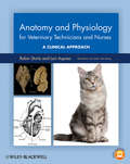
Anatomy and Physiology for Veterinary Technicians and Nurses
by Lori Asprea Robin SturtzAnatomy and Physiology for Veterinary Technicians and Nurses: A Clinical Approach is a comprehensive resource on the anatomy and physiology of dogs and cats, with comparisons to horses, birds, and ruminants. Organized by body system with a comparative approach, the book follows a unique format by addressing anatomy separately from physiology for clarity and improved comprehension. Each anatomy chapter has a corresponding physiology chapter, complete with illustrations, charts, and boxes to promote understanding.Written specifically for veterinary technicians and nurses, the book applies anatomy and physiology to clinical practice, with case examples demonstrating clinical relevance. The figures from the book, additional questions and answers, labeling quizzes, teaching PowerPoints, and a dissection video are available online at www.wiley.com/go/sturtz. This introduction to body system analysis of normal structure and function is a must-have resource for students of veterinary technology and nursing, as well as a useful quick review for the busy professional.
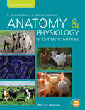
Anatomy and Physiology of Domestic Animals
by D. Michael Denbow R. Michael AkersAnatomy and Physiology of Domestic Animals, Second Edition offers a detailed introduction to the foundations of anatomy and physiology in a wide range of domestic species. Well illustrated throughout, the book provides in-depth information on the guiding principles of this key area of study for animal science students, fostering a thorough understanding of the complex make-up of domestic animals. This Second Edition includes access to supplementary material online, including images and tables available for download in PowerPoint, a test bank of questions for instructors, and self-study questions for students at www.wiley.com/go/akers/anatomy.Taking a logical systems-based approach, this new edition is fully updated and now provides more practical information, with descriptions of anatomic or physiological events in pets or domestic animals to demonstrate everyday applications. Offering greater depth of information than other books in this area, Anatomy and Physiology of Domestic Animals is an invaluable textbook for animal science students and professionals in this area.
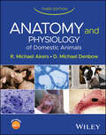
Anatomy and Physiology of Domestic Animals
by D. Michael Denbow R. Michael AkersComprehensive resource on the anatomy and physiology systems of common domestic animals, with learning resources included throughout Anatomy and Physiology of Domestic Animals bridges the gap between theory and practice, emphasizing real-world applications. In this newly revised and updated Third Edition, each chapter includes a short section which emphasizes current animal management practices that take advantage of physiological principles discussed in that chapter to improve animal growth, development, or function. Instructors will gain access to a website with PowerPoint slides of all of the figures, tables, and illustrations used in the book, with one PowerPoint presentation for each chapter. A test bank of potential questions for each book chapter is featured, including short answer, matching, true and false, and discussion questions. Each chapter also includes a study guide located at the end of each chapter and an opening section that provides an outline and listing of key concepts that the reader should get from each chapter. Some of the key revisions to this Third Edition of Anatomy and Physiology of Domestic Animals include: Genetic testing and modification of DNA to improve animal health or performance and the use of RNA to create vaccinesThe dynamic nature of skin, not just as physical protection, but also in its relevance in immunityThe role of supportive non-neurons and proteins in brain functionNew discoveries in hormone signaling and uses of hormone therapies in domestic animalsReproductive strategies to regulate estrus, breeding schemes, and sex of offspring Anatomy and Physiology of Domestic Animals is an essential up-to-date reference for undergraduate students in animal science, dairy science, pre-veterinary medicine, veterinary technician training, and biology. The book is also relevant as reference/review text for graduate students in animal sciences and physiology.
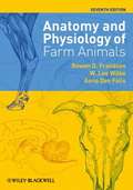
Anatomy and Physiology of Farm Animals
by Anna Dee Fails Rowen D. Frandson W. Lee WilkeThe Seventh Edition of Anatomy and Physiology of Farm Animals is a thoroughly updated and revised version of this classic text. Drawing on current science and terminology with a number of new illustrations throughout and a new chapter on poultry, the book maintains its reputation for clarity, balanced scope, and breadth of content. The Seventh Edition provides veterinary, animal science, agriculture, and veterinary technician students with a comprehensive yet clear reference to understanding the fundamentals of anatomy and physiology.
Anatomy and Physiology of Farm Animals
by Anna Dee Fails Christianne MageeRevised and updated, the eighth edition of Anatomy and Physiology of Farm Animals remains the essential resource for detailed information on farm animal anatomy and physiology. Offers a revised edition to this comprehensive guide to the anatomy and physiology of farm animals Presents learning objectives in each chapter for the first time Adds new material on endocrine and metabolic regulation of growth and body composition Features additional illustrations to enhance comprehension Includes a companion website that offers supplemental content, including word roots, clinical cases, study and practice questions, the images from the book and additional images, diagrams, and videos to enhance learning.
Anatomy and Physiology of Farm Animals
by Anna Dee Fails Christianne MageeA complete guide to the anatomy and physiology of farm animals, fully updated and revised In the newly revised ninth edition of Anatomy and Physiology of Farm Animals, distinguished veterinary professors Drs. Anna Fails and Christianne Magee deliver a comprehensive guide for animal science, veterinary technician, and pre-veterinary students and instructors seeking a well-organized and easy-to-understand resource. The new edition offers modified and refined learning objectives at the beginning of each chapter, as well as a brand-new chapter on llamas/alpacas that highlights the significant species differences and explains the roles of these species in the wool and packing industries. Additional illustrations enhance comprehension and improve the anatomy sections of the book. New “Study Prompts,” integrative application questions, are included in each chapter in differently colored text and stimulate understanding of the material. Finally, a reorganized companion website is included with the book. It integrates fully with the print text and provides supplemental content, including word roots, clinical cases, study and practice questions, and additional images, diagrams, and videos. Readers will also find: An excellent anatomy and physiology resource for high school and undergraduate students in animal science, veterinary medicine, and zoology programsComprehensive explorations of the anatomy and physiology of the cell Practical discussions of embryology, the skeletal system, and microscopic anatomy Complete discussion of the physiology of muscle and the anatomy and physiology of the nervous system A valuable comprehensive resource for advanced high school and undergraduate animal science students in agriculture, pre-veterinary, and veterinary technical program, Anatomy and Physiology of Farm Animals will also benefit people practicing in allied professions and veterinary practitioners.
Anatomy and Physiology of Hearing for Audiologists
by Kevin K. Ohlemiller William W. ClarkThe first anatomy and physiology text just for audiologists, this new text brings together some of the best professional minds in the field to consider the structures and mechanisms of the auditory system. <P><P>Basic science is covered in the foundations section of the text, giving a much needed examination of the biological processes in terms the audiologist needs most. <P><P>Detailed examination of the anatomy and physiology of hearing follows with diagrams and in-depth discussions. The text concludes with chapters on the pathology of hearing, covering the different causes of hearing loss, from noise-induced hearing loss to genetic aspects of hearing loss. <P><P>From start to finish this text is written specifically for the audiologist, making it an essential foundational resource.

Anatomy and Physiology of the Circulatory and Ventilatory Systems (Biomathematical and Biomechanical Modeling of the Circulatory and Ventilatory Systems #6)
by Marc ThirietTogether, the volumes in this series present all of the data needed at various length scales for a multidisciplinary approach to modeling and simulation of flows in the cardiovascular and ventilatory systems, especially multiscale modeling and coupled simulations. The cardiovascular and respiratory systems are tightly coupled, as their primary function is to supply oxygen to, and remove carbon dioxide from, the body's cells. Because physiological conduits have deformable and reactive walls, macroscopic flow behavior and prediction must be coupled to nano- and microscopic events in a corrector scheme of regulated mechanism. Therefore, investigation of flows of blood and air in physiological conduits requires an understanding of the biology, chemistry, and physics of these systems, together with the mathematical tools to describe their functioning in quantitative terms. The present volume focuses on macroscopic aspects of the cardiovascular and respiratory systems in normal conditions, i. e. , anatomy and physiology, as well as the acquisition and processing of medical images and physiological signals.
Anatomy and Physiology: An Integrative Approach
by Michael P. McKinley Valerie Dean O'Loughlin Theresa Stouter BidleHuman anatomy and physiology is a fascinating subject. However, students can be overwhelmed by the complexity, the interrelatedness of concepts from different chapters, and the massive amount of material in the course. Our goal was to create a textbook to guide students on a clearly written and expertly illustrated beginner's path through the human body.
Anatomy and Physiology: The Unity of Form and Function (6th Edition)
by Kenneth S. SaladinWith Saladin, students make connections through learning outcomes and assessments, integrated media, and a writing style that clearly depicts anatomy and physiology processes. A consistent set of chapter learning tools helps students identify and retain key concepts while the stunning visual program provides a realistic view of body structures and processes. Saladin's text requires no prior knowledge of college chemistry or cell biology, and is designed for a two-semester A&P course.
Anatomy and the Organization of Knowledge, 1500–1850 ("The Body, Gender and Culture" #9)
by Matthew Landers Brian MuñozAcross early modern Europe, the growing scientific practice of dissection prompted new and insightful ideas about the human body. This collection of essays explores the impact of anatomical knowledge on wider issues of learning and culture.
Anatomy at a Glance
by Simon Blackburn Omar Faiz David MoffatFollowing the familiar, easy-to-use at a Glance format, and in full-colour, this new edition provides an accessible introduction and revision aid for medical, nursing and all health sciences students. Thoroughly updated and now fully supported by a set of web-based flashcards, Anatomy at a Glance provides a user-friendly overview of anatomy to encapsulate all that the student needs to know. Anatomy at a Glance: Addresses the basic concepts of anatomy in an highly visual, easy-to-remember way Features two new chapters outlining anatomical terminology and basic embryology Includes more coverage of imaging techniques such as CT and MRI Offers free online flashcards for self-assessment and revision at www. wiley. com/go/anatomyataglance To find out more about the at a Glance series, please visit www. ataglanceseries. com
Anatomy for Anaesthetists
by Andrew Lawson Harold EllisFirst published in 1963, Anatomy for Anaesthesists is the definitive anatomy text for anaesthetists in training and remains an invaluable reference for those in practice. The text explores in depth those areas of particular interest to anaesthetists: the respiratory pathway, the heart, the vertebral canal and its contents, the peripheral nerves, the autonomic nervous system, and the cranial nerves, and also includes sections on the anatomy of pain and other zones of anaesthetic interest.This new 9th edition has been fully revised and updated to incorporate developments in regional techniques and the increased use of ultrasound.