- Table View
- List View
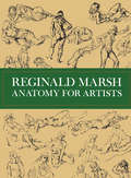
Anatomy for Artists (Dover Anatomy for Artists)
by Reginald MarshPortraying the living human form, not only with anatomical accuracy, but so that it conveys motion, emotion, and vitality is one of the greatest challenges faced by the artist. In the studies in this volume, famous artist and art instructor Reginald Marsh brought his genius to bear on the complex problem of life drawing. Delving into the work of the great masters (Michelangelo, Leonardo, Raphael, Rubens, Poussin, Dürer, Holbein, and others), Marsh simplified, abstracted, adapted, and reinterpreted their work into a collection of drawings both immensely interesting and instructive to the practicing artist and the student. The 209 pages of drawings in this volume show the human body in a wide variety of positions, viewed from many different angles. Marsh directs special attention to those angles, aspects, and physical positions which are the most difficult to portray. His great talent, coupled with a rare ability to instruct others (Marsh taught at the Art Students League for many years) gave him unusual sensitivity to the concerns of the artist in life drawing: his concise commentary on the drawing points up the problems addressed in each -- tone, movement, proportion, composition, etc. The front, side, back, head, arms and hands, legs and feet, and full figure drawings are all included. A separate section on the problems of proportion explores 7, 7 1/2, and 8 head schemes, providing an unusually workable and lucid treatment of the topic for the practicing artist. The body and parts of the body are drawn in skeleton, tissue and muscle, major bone structure, and as they appear in life. Marsh studied medical anatomy as well as the work of the great medical artists in order to perfect his knowledge of human anatomy. All of the hundreds of drawings, figures, and details of this volume have been excellently reproduced in this edition. The last 95 drawings in the book are all original studies by Marsh, never before published in book form. These casual, light-hearted drawings (mostly of female nudes) illustrate both Marsh's seemingly easy mastery of the techniques of life drawing, and his characteristic lusty, Rubenesque style. Because they are so distinctly in his own style, these drawings highlight the great scope and knowledgeability he has shown in the earlier instructive studies. Those who know and admire Marsh as an artist, as well as anyone who wishes to learn to draw from life, will find this volume indispensable.
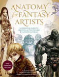
Anatomy for Fantasy Artists: An Essential Guide to Creating Action Figures & Fantastical Forms
by Glenn FabryAn indispensable guide for anyone interested in improving and developing their fantasy art figures. Start with the basics of human anatomical drawing and musculature, and then learn ways to distort, develop, and transform the human figure, giving it features that range from monstrous or magical to super-agile or larger than life.
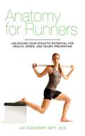
Anatomy for Runners: Unlocking Your Athletic Potential for Health, Speed, and Injury Prevention
by Jay DicharryRunning has become more and more popular in recent years, with thousands of people entering marathons, buying new running shoes with the latest technology, and going for a daily jog, whether on the track or on a treadmill. Unfortunately, with running comes injuries, as a result of wrong information and improper training. Author Jay Dicharry was tired of getting the same treatments from doctors that didn't heal his joint and muscle pain from running, so he decided to combine different fields of clinical care, biomechanical analysis, and coaching to help you avoid common injuries and become the best runner you can be. Along with clear and thorough explanations of how running influences the body, and how the body influences your running, this book answers many of the common questions that athletes have: Do runners need to stretch? What is the best way to run? What causes injuries? Which shoes are best for running? Is running barefoot beneficial? The mobility and stability tests will assess your form, and the corrective exercises, along with step-by-step photos, will improve your core and overall performance, so that you can train and run with confidence, knowing how to avoid injuries!
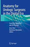
Anatomy for Urologic Surgeons in the Digital Era: Scanning, Modelling and 3D Printing
by Emre Huri Domenico VenezianoThis book provides a practical guide in the use of imaging and visualization technologies in urology. It details how output from diagnostic systems, can be represented through synthetic, virtual and augmented reality tools, such as holograms and three dimensional (3D) modelling and how they can improve everyday surgical procedures including laparoscopic, robotic-assisted, open, endoscopic along with the latest and most innovative approaches. Anatomy for Urologic Surgeons in the Digital Era: Scanning, Modelling and 3D Printing systematically reviews diagnostic imaging, visualization tools available in urology and is a valuable resource for all practicing and in-training urological surgeons.
Anatomy for the FRCA
by James Bowness Alasdair TaylorAnatomy questions are asked in all parts of the FRCA examinations, and for many trainees it is a particularly daunting part of the exams. This important new book provides a comprehensive, exam-orientated clinical anatomy book for anaesthetists preparing for all parts of the FRCA. It covers all body regions, relating underlying anatomy to practical procedures and anatomical principles, spanning the breadth of the curriculum and comprising exam-style questions: a chapter of SAQ questions, one of OSCE stations, one of SOE questions and one of MCQs. The text is highly illustrated in full colour with ultrasound images, diagrams and photographs of cadaveric material and models. This is the first anatomy book specifically orientated for the FRCA exam, making it an essential resource for anaesthetists preparing for all parts of the FRCA examination.
Anatomy in Action: The Dynamic Muscular Systems that Create and Sustain the Moving Body
by Theodore DimonAn illustrated guide to the core design principles of the body&’s musculoskeletal system—for kinesiologists, movement therapists, yoga teachers, dancers, and bodyworkers of all kindsWhat does knowledge of anatomical structure have to do with preventing everyday muscular aches, pains, and injuries? According to Dr. Theodore Dimon, everything!Our bodies are designed to work holistically, supported by an intelligently organized system of muscles, bones, and connective tissue. So when we target problem spots by stretching, relaxing, or strengthening individual muscles, we bypass the dynamic, interconnected network that enables healthy functioning and injury prevention. Understanding how this system works in action is the key.In this groundbreaking guide, Dr. Dimon describes the basic principles that govern our bodies&’ musculoskeletal architecture and provides practical exercises to activate specific muscle groups and demonstrate our bodies&’ efficient holistic function. Readers will learn about dynamic design and the body in action, including: • How the musculoskeletal system works as a whole • The relationship between proprioception and muscle length • About maximizing spinal, shoulder, hip, arm, and leg stability and health • The important role of breath and breathing • About posture and musculoskeletal support With more than 300 illustrations, this is an ideal resource for students and practitioners of kinesiology, bodywork, movement, sport kinesiology, dance, and all readers searching for a dynamic guide to the human body.
Anatomy in Diagnostic Imaging
by J?rgen Tranum-Jensen Peter FleckensteinNow in its third edition, Anatomy in Diagnostic Imaging is an unrivalled atlas of anatomy applied to diagnostic imaging. The book covers the entire human body and employs all the imaging modalities used in clinical practice; x-ray, CT, MR, PET, ultrasound and scintigraphy. An introductory chapter explains succinctly the essentials of the imaging and examination techniques drawing on the latest technical developments.In view of the great strides that have been made in this area recently, all chapters have been thoroughly revised in this third edition. The book's original and didactically convincing presentation has been enhanced with over 250 new images. There are now more than 900 images, all carefully selected in order to be user-friendly and easy-to-read, due to their high quality and the comprehensive anatomical interpretation directly placed alongside every one.Both for medical students and practising doctors, Anatomy in Diagnostic Imaging, Third edition will serve as the go-to all-round reference collection linking anatomy and modern diagnostic imaging.
Anatomy of Anorexia
by Steven Levenkron"Invaluable to clinicians, parents, teenagers, and adults who are struggling with anorexia." —Lynn E. Ponton, M.D. Anatomy of Anorexia is a tremendous tool for families: now more than ever, early diagnosis and treatment, and family participation, are crucial in helping the anorexic. Preeminent therapist Steven Levenkron demystifies this life-threatening disease and shows how the millions of girls and women who are afflicted with anorexia can be helped—and can look forward to rich and productive lives. "The nation’s premier expert in treating anorexia has written the nation’s premier book for parents, relatives, and friends of young women afflicted with this life-threatening disease."—Joseph A. Califano Jr., president of the National Center on Addiction and Substance Abuse at Columbia University and former U.S. Secretary of Health, Education, and Welfare "[Levenkron’s] insights, descriptions of family relationships, and treatment recommendations for therapists create a rich, deep, and most helpful guide for a community of people whose lives are deeply and painfully affected by this persistent illness."—Samuel C. Klagsbrun, M.D.

Anatomy of Authoritarianism in the Arab Republics
by Joseph SassoonBy examining the system of authoritarianism in eight Arab republics, Joseph Sassoon portrays life under these regimes and explores the mechanisms underpinning their resilience. How did the leadership in these countries create such enduring systems? What was the economic system that prolonged the regimes' longevity, but simultaneously led to their collapse? Why did these seemingly stable regimes begin to falter? This book seeks to answer these questions by utilizing the Iraqi archives and memoirs of those who were embedded in these republics: political leaders, ministers, generals, security agency chiefs, party members, and business people. Taking a thematic approach, the book begins in 1952 with the Egyptian Revolution and ends with the Arab uprisings of 2011. It seeks to deepen our understanding of the authoritarianism and coercive systems that prevailed in these countries and the difficult process of transition from authoritarianism that began after 2011.
Anatomy of Cranial Arteries, Embryology and Variants
by Thomas Robert Sara Bonasia Michel W. BojanowskiThis book on the anatomy of central nervous system arteries concentrates on all anatomical variations of the central nervous system and it describes the embryological processes that hide behind the possible adult variants.The first section of the work is a reminder of general concepts of embryology. After that, each section corresponds to arteries of an anatomical location: intradural, dural, skull base and cranio-cervical junction. Each chapter is dedicated to a single artery to facilitate the reader’s search for information. In addition, modern and detailed illustrations of the embryological steps and adult variants are included. There are two types of illustrations: artist’s drawing, usually to explain the vascular embryology, and angiographic images. The central point of the book lies in the space devoted to the embryological development of each artery and the processes that can lead to the development of different variants in the adult. The audience of this book is aimed at neurosurgeons and neuroradiologists, specialists in the neurovascular area, but it will also help residents in neurosurgery, neuroradiology and neurology in their daily practice.
Anatomy of Development: Concept, Theory, and Practice
by Bikram PattanaikThis book details the fundamentals of development studies by adopting a multi-disciplinary approach. It presents a balanced mix of economic, social, political, cultural and administrative premises of development and analyzes its theoretical and practical dimensions. It also provides insight into the role of the stakeholders of development in different sectors.The volume provides a holistic understanding of development, effectively demonstrating how it differs from economic growth. Beginning with development theories, paradigms and actors involved in the development process, this book goes on to explain the concepts of development administration, development governance, development planning, development management and development communication. One of the fundamental components of the book is the elucidation of Development Theories – classical, neoclassical, developmental and heterodox theories essential to the discipline of “development”.This book will be useful to undergraduate and postgraduate students, researchers, teachers of development studies, economics, sociology, political science, and public administration. It will also be useful to administrators and development administration officials of state and central governments, planners, policymakers and people working in NGOs, in addition to corporate sector functionaries dealing with corporate social responsibilities and those handling developmental issues and challenges.
Anatomy of Flowering Plants: An Introduction to Plant Structure and Development
by Paula J. RudallUnderstanding plant anatomy is not only fundamental to the study of plant systematics and palaeobotany, but is also an essential part of evolutionary biology, physiology, ecology and the rapidly expanding science of developmental genetics. This modernised new edition covers all aspects of comparative plant structure and development, arranged in a series of chapters on the stem, root, leaf, flower, pollen, seed and fruit. Internal structures are described using magnification aids from the simple hand-lens to the electron microscope. Numerous references to recent topical literature are included, and new illustrations reflect a wide range of flowering plant species. The phylogenetic context of plant names has been updated as a result of improved understanding of the relationships among flowering plants. This clearly written text is ideal for students studying a wide range of courses in botany and plant science, and is also an excellent resource for professional and amateur horticulturists.
Anatomy of Four Race Riots: Racial Conflict in Knoxville, Elaine (Arkansas), Tulsa, and Chicago, 1919-1921
by Lee E. Williams Lee E. Williams IIAnatomy of Four Race Riots is a study of the terrible racial violence that erupted in four different communities of America during the post World War I years, racial violence that left hundreds dead or injured and a massive amount of destruction in its wake.Although the igniting incident or event varied somewhat, there was a similarity in the racial climate that existed in each town. The emerging blacks, boosted economically and idealistically by the war effort, were viewed as a threat by some of the whites. The bloody confrontations described here were grave evidence of the intensity of the fear and hatred that existed between a portion of the races.
Anatomy of Game Design
by Tom SmithPeople have played games forever, but it’s only in the past few decades that people really started thinking about what games are, how they work, and how to make them better.Anatomy of Game Design takes some of the most popular and beloved games of all time and dissects them to see what makes them tick. By breaking down the systems and content of each game, the underlying systems of game design are laid bare.Eight games are analyzed – including Settlers of Catan; Centipede; Candy Crush Saga; Papers, Please; Magic: The Gathering; and more – each representing a different genre or era of game design. Each game is discussed in detail, using the same methods for each game. What are the verbs of the game that give the player agency? How do those verbs fit together to form a core loop that makes the game engaging? What are the systems that power the gameplay? What is the larger flow that makes the game interesting over and over again?Each game is then used as an example to tie back to one or more larger topics in game design, such as systems design, randomness, monetization, game theory, and iterative approaches to game development.Key Features: Uses well-known games to provide specific, discrete examples of broader game design theory Discusses eight popular games using the same methodology to allow comparison of different types of games Includes both high-level theory and academic perspective and practical, real-world guidance from a working game designer who has created these games for commercial release Provides clear direction for deeper inquiry into game design or related fields such as psychology, anthropology, game development, or systems thinking
Anatomy of Global Stock Market Crashes: An Empirical Analysis (SpringerBriefs in Economics)
by Chitrakalpa Sen Gagari ChakrabartiThis work is an exploration of the global market dynamics, their intrinsic natures, common trends and dynamic interlinkages during the stock market crises over the last twelve years. The study isolates different phases of crisis and differentiates between any crisis that remains confined to the region and those that take up a global dimension. The latent structure of the global stock market, the inter-regional and intra-regional stock market dynamics around the crises are analyzed to get a complete picture of the structure of the global stock market. The study further probing into the inherent nature of the global stock market in generating crisis finds the global market to be chaotic thus making the system intrinsically unstable or at best to follow knife-edge stability. The findings have significant bearing at theoretical level and on policy decisions.
Anatomy of Hard Talks: What Makes a Conversation Fail
by Holly WeeksEvery difficult conversation is unique, but what separates hard talks from normal conversations is not as individual as we might think. Across the board, there are three traits - with real costs - that make difficult conversations categorically different: combat mentality, emotional loads, and being hard to read. In addition, our simple and familiar ideas about how to handle tough conversations don't help us. We can't get through these conversations if we don't look more closely at what we actually do that makes them go bad.
Anatomy of Injustice: A Murder Case Gone Wrong
by Raymond BonnerThe book that helped free an innocent man who had spent twenty-seven years on death row. In January 1982, an elderly white widow was found brutally murdered in the small town of Greenwood, South Carolina. Police immediately arrested Edward Lee Elmore, a semiliterate, mentally retarded black man with no previous felony record. His only connection to the victim was having cleaned her gutters and windows, but barely ninety days after the victim’s body was found, he was tried, convicted, and sentenced to death. Elmore had been on death row for eleven years when a young attorney named Diana Holt first learned of his case. After attending the University of Texas School of Law, Holt was eager to help the disenfranchised and voiceless; she herself had been a childhood victim of abuse. It required little scrutiny for Holt to discern that Elmore’s case-plagued by incompetent court-appointed defense attorneys, a virulent prosecution, and both misplaced and contaminated evidence-reeked of injustice. It was the cause of a lifetime for the spirited, hardworking lawyer. Holt would spend more than a decade fighting on Elmore’s behalf. With the exemplary moral commitment and tenacious investigation that have distinguished his reporting career, Bonner follows Holt’s battle to save Elmore’s life and shows us how his case is a textbook example of what can go wrong in the American justice system. He reviews police work, evidence gathering, jury selection, work of court-appointed lawyers, latitude of judges, iniquities in the law, prison informants, and the appeals process. Throughout, the actions and motivations of both unlikely heroes and shameful villains in our justice system are vividly revealed. Moving, suspenseful, and enlightening,Anatomy of Injusticeis a vital contribution to our nation’s ongoing, increasingly important debate about inequality and the death penalty.
Anatomy of Innocence: Testimonies of the Wrongfully Convicted
by Barry Scheck Scott Turow Laura Caldwell Leslie S. KlingerRecalling the great muckrakers of the past, an outraged team of America’s best-selling writers unite to confront the disasters of wrongful convictions. Wrongful convictions, long regarded as statistical anomalies in an otherwise sound justice system, now appear with frightening regularity. But few people understand just how or why they happen and, more important, the immeasurable consequences that often haunt the lucky few who are acquitted, years after they are proven innocent. Now, in this groundbreaking anthology, fourteen exonerated inmates narrate their stories to a roster of high-profile mystery and thriller writers—including Lee Child, Sara Paretsky, Laurie R. King, Jan Burke and S. J. Rozan—while another exoneree’s case is explored in a previously unpublished essay by legendary playwright Arthur Miller. An astonishing and unique collaboration, these testimonies bear witness to the incredible stories of innocent men and women who were convicted of serious crimes and cast into the maw of a vast and deeply flawed American criminal justice system before eventually, and miraculously, being exonerated. Introduced by best-selling authors Scott Turow and Barry Scheck, these master storytellers capture the tragedy of wrongful convictions as never before and challenge readers to confront the limitations and harsh realities of the American criminal justice system. Lee Child tells of Kirk Bloodsworth, who obsessively read about the burgeoning field of DNA testing, cautiously hoping that it held the key to his acquittal—until he eventually became the first person to be exonerated from death row based on DNA evidence. Judge John Sheldon and author Gayle Lynds team up to share Audrey Edmunds’s experience raising her children long distance from her prison cell. And exoneree Gloria Killian recounts to S. J. Rozan her journey from that fateful "knock on the door" and the initial shock of accusation to the scars she carries today. Together, the powerful stories collected within the Anatomy of Innocence detail every aspect of the experience of wrongful conviction, as well as the remarkable depths of endurance sustained by each exoneree who never lost hope.
Anatomy of Japanese Business
by Kasuo SatoThis volume collects eleven essays written by Japanese experts on various aspects of Japanese business management and is a sequel to the volume Industry and Business in Japan. It examines the mechanisms for Japan 's phenomenal economic growth since the Second World War by analyzing Japanese management, business groups, production systems and business strategy.
Anatomy of Love: A Natural History of Mating, Marriage, and Why We Stray (Completely Revised and Updated with a New Introduction)
by Helen FisherA contemporary classic about love now completely revised and updated. First published in 1992, Helen Fisher’s “fascinating” (New York Times) Anatomy of Love quickly became a classic. Since then, Fisher has conducted pioneering brain research on lust, romantic love, and attachment; gathered data on more than 80,000 people to explain why you love who you love; and collected information on more than 30,000 men and women on sexting, hooking up, friends with benefits, and other current trends in courtship and marriage. And she presents a new, scientifically based and optimistic perspective on relationships in our digital age—what she calls “slow love.” This is a cutting-edge tour de force that traces human family life from its origins in Africa over 20 million years ago to the Internet dating sites and bedrooms of today. And it’s got it all: the copulatory gaze and other natural courting ploys; the who, when, where, and why of adultery; love addictions; her discovery of four broad chemically based personality styles and what each seeks in romance; the newest data on worldwide (biologically based) patterns of divorce; how and why men and women think differently; the real story of women, men, and power; the rise—and fall—of the sexual double standard; and what brain science tells us about how to make and keep a happy partnership.
Anatomy of Malice: The Enigma of the Nazi War Criminals
by Joel E. DimsdaleAn eminent psychiatrist delves into the minds of Nazi leadershipin &“a fresh look at the nature of wickedness, and at our attempts to explain it&” (Sir Simon Wessely, Royal College of Psychiatrists). When the ashes had settled after World War II and the Allies convened an international war crimes trial in Nuremberg, a psychiatrist, Douglas Kelley, and a psychologist, Gustave Gilbert, tried to fathom the psychology of the Nazi leaders, using extensive psychiatric interviews, IQ tests, and Rorschach inkblot tests. The findings were so disconcerting that portions of the data were hidden away for decades and the research became a topic for vituperative disputes. Gilbert thought that the war criminals&’ malice stemmed from depraved psychopathology. Kelley viewed them as morally flawed, ordinary men who were creatures of their environment. Who was right? Drawing on his decades of experience as a psychiatrist and the dramatic advances within psychiatry, psychology, and neuroscience since Nuremberg, Joel E. Dimsdale looks anew at the findings and examines in detail four of the war criminals, Robert Ley, Hermann Göring, Julius Streicher, and Rudolf Hess. Using increasingly precise diagnostic tools, he discovers a remarkably broad spectrum of pathology. Anatomy of Malice takes us on a complex and troubling quest to make sense of the most extreme evil. &“In this fascinating and compelling journey . . . a respected scientist who has long studied the Holocaust asks probing questions about the nature of malice. I could not put this book down.&”—Thomas N. Wise, MD, Johns Hopkins University School of Medicine &“This harrowing tale and detective story asks whether the Nazi War Criminals were fundamentally like other people, or fundamentally different.&”—T.M. Luhrmann, author of How God Becomes Real
Anatomy of Medical School Admissions: Need-to-Know Information about Getting into Med School, the MCAT, and What it Takes to Be a Doctor (Graduate School Admissions Guides)
by Princeton ReviewThe Princeton Review Knows Med School. Thinking about a career in medicine? This is the book you need to ensure you're prepared in every way: Learn about medical school admissions, career options, how to prepare yourself as an undergrad, and, of course, about the major changes coming to the MCAT in 2015. The health system of tomorrow will require a different kind of physician. With over 30 years of experience and a proven track record helping millions of students achieve their goals, The Princeton Review is the only company with the expertise and resources to guide you through each step of your journey. We're not just about test prep: we're about prepping for your life. Inside this eBook, you'll find guidance and advice on topics like:* Do You Have What it Takes? (Thinking Like a Medical Student)* Is a Career in Medicine Right For You? (What They Don't Tell You at Career Day)* Everything You Need to Know about Med School Admissions* Researching Medical Schools, or ... Making the List* Q&A With Admissions Officers* The Inside Scoop on Applications, Personal Statements, Financial Aid, and More ...* How to Rock the Interview* The MCAT: Now and 2015* Preparing for and Taking the MCAT* Sample Personal Statements
Anatomy of Orofacial Structures (Seventh Edition)
by Richard W. Brand Donald E. IsselhardThis comprehensive edition on orofacial structures provides a complete introduction with dedicated sections on dental anatomy,oral histology and embryology, head and neck anatomy.

Anatomy of Paradise Hawaii and the Islands of the South Seas
by J. C. FurnasEmbark on an enchanting journey through the idyllic islands of the Pacific with J. C. Furnas's Anatomy of Paradise: Hawaii and the Islands of the South Seas. This captivating travel narrative offers a rich and detailed exploration of the cultures, landscapes, and histories of Hawaii and the South Sea Islands, capturing the essence of these paradisiacal destinations.Furnas, a skilled writer and keen observer, provides readers with a comprehensive account of his travels through some of the world's most breathtaking and remote locales. From the volcanic majesty of Hawaii to the serene beauty of Tahiti and Fiji, Anatomy of Paradise vividly portrays the natural splendor and unique cultural heritage of each island.The book delves into the complex history of the Pacific Islands, tracing their journey from ancient Polynesian navigators to encounters with European explorers and the impact of colonialism. Anatomy of Paradise is not just a travelogue; it is an exploration of the human spirit and its connection to these enchanting lands. Furnas highlights the resilience, traditions, and daily lives of the islanders, providing a respectful and empathetic portrayal of their societies. His engaging prose brings to life the rhythms of island life, from traditional ceremonies and dances to the challenges of modernity.This book is an essential read for travel enthusiasts, historians, and anyone fascinated by the Pacific Islands. Furnas's evocative storytelling and thorough research create a vivid tapestry of the islands, making Anatomy of Paradise a timeless tribute to one of the world's most captivating regions.Join J. C. Furnas on this unforgettable journey and experience the magic, mystery, and beauty of Hawaii and the Islands of the South Seas through the eyes of a masterful storyteller.
Anatomy of Regional Disparities in the Slovak Republic
by Mariusz Jarmuzek Biswajit BanerjeeA report from the International Monetary Fund.