- Table View
- List View
ICP Emissionsspektrometrie für Praktiker
by Joachim NölteDas Basis-Know-how für richtige ICP-OES-Analytik!Erstmalig ist eine deutschsprachige, leicht verständliche und anwenderorientierte Einführung in die ICP-Emissionspektrometrie verfügbar. Sie umfaßt die praxisrelevanten Grundlagen, gerätetechnische Informationen, eine Anleitung zur Methodenentwicklung und viele praktische Anwendungsbeispiele. Das Buch ist kompakt und sehr übersichtlich gestaltet, mit Infoboxen zu typischen Fragen und Problemen, Checklisten und detaillierten Hinweisen zur Handhabung. Es ist nicht nur ein Begleiter für die eigenständige Aus- und Weiterbildung, sondern ebenso ein verlässlicher Leitfaden für die praktische Laborarbeit, denn auch die Aspekte Pflege und Wartung sowie Trouble-Shooting kommen nicht zu kurz.Alle Anwender der ICP-OES können vom bewährten Erfahrungsschatz des Autors profitieren, den er in zwei Jahrzehnten bei der Ausbildung und Beratung von Anwendern sowie bei der Geräteentwicklung gesammelt hat. Er war Mitarbeiter eines führenden Geräteherstellers und ist jetzt freiberuflicher Berater.
ICPES 2019: Selected articles from the 9th International Conference on Power and Energy Systems, Perth, Australia (Lecture Notes in Electrical Engineering #669)
by Farhad Shahnia Sara DeilamiThis book highlights various applications of renewable energy systems and their enabling technologies in electrical power systems. It features selected articles from the 9th International Conference on Power and Energy Systems (ICPES 2019), held in Perth, Australia, which presented the latest advances in the field and provided a platform to exchange ideas and foster future collaboration with a sustainable future in mind.
ICREEC 2019: Proceedings of the 1st International Conference on Renewable Energy and Energy Conversion (Springer Proceedings in Energy)
by Ahmed Belasri Sid Ahmed BeldjilaliThis book highlights peer reviewed articles from the 1st International Conference on Renewable Energy and Energy Conversion, ICREEC 2019, held at Oran in Algeria. It presents recent advances, brings together researchers and professionals in the area and presents a platform to exchange ideas and establish opportunities for a sustainable future. Topics covered in this proceedings, but not limited to, are photovoltaic systems, bioenergy, laser and plasma technology, fluid and flow for energy, software for energy and impact of energy on the environment.
ICRRM 2019 – System Reliability, Quality Control, Safety, Maintenance and Management: Applications to Civil, Mechanical and Chemical Engineering
by Vinit Kumar Gunjan Sri Niwas Singh Tran Duc-Tan Gloria Jeanette Rincon Aponte Amit KumarContent of this proceedings discusses emerging trends in structural reliability, safety and disaster management, covering topics like total quality management, risk maintenance and design for reliability. Some papers also address chemical process reliability, reliability analysis and engineering applications in chemical process equipment systems and includes a chapter on reliability evaluation models of chemical systems. Accepted papers from 2019 International Conference on Reliability, Risk Maintenance and Engineering Management (ICRRM 2019) are part of this conference proceeding. It offers useful insights to road safety engineers, disaster management professionals involved in product design and probabilistic methods in manufacturing systems.
ICSBE 2018: Proceedings of the 9th International Conference on Sustainable Built Environment (Lecture Notes in Civil Engineering #44)
by Ranjith Dissanayake Priyan MendisThis book highlights current research and development in the area of sustainable built environments, currently one of the most important disciplines in civil engineering. It covers a range of topics, including sustainable construction and infrastructures, waste and wastewater management, enhanced sustainability, renewable and clean energy, sustainable materials and industrial ecology, building automation and virtual reality, and impact of climate change. As such it provides vital insights into responsible urbanization practices, and new tools and technologies in civil engineering that can mitigate the negative effects of the built environment.
ICSBE 2022: Proceedings of the 13th International Conference on Sustainable Built Environment (Lecture Notes in Civil Engineering #362)
by Ranjith Dissanayake Priyan Mendis Kolita Weerasekera Sudhira De Silva Shiromal Fernando Chaminda Konthesingha Pradeep GajanayakeThis book highlights the latest knowledge and innovations in the fields of civil engineering and construction industry striving for a sustainable built environment. It consists of high quality and innovative research findings selected from the proceedings of the 13th ICSBE 2022 under the themes of sustainable construction, urban green infrastructure and planning, rainwater harvesting and water conservation, high-performance concrete, indoor environmental quality and indoor plants, wind and hydro-power energy, waste and wastewater management for enhanced sustainability, impacts of climate change, carbon footprint, global climate model and landscaping, material flows and industrial ecology, sustainable materials, etc.
ICSCEA 2019: Proceedings of the International Conference on Sustainable Civil Engineering and Architecture (Lecture Notes in Civil Engineering #80)
by J. N. Reddy Chien Ming Wang Van Hai Luong Anh Tuan LeThis book presents papers from the International Conference on Sustainable Civil Engineering and Architecture 2019, which was held in Ho Chi Minh City, Vietnam, from 24–26 October 2019. The conference brought together international experts from both academia and industry to share their knowledge and experiences, and to facilitate collaboration and improve cooperation in the field. The book highlights the latest advances in sustainable architecture and civil engineering, covering topics such as offshore structures, structural engineering, construction materials, and architecture.
ICSDEMS 2019: Proceedings of the International Conference on Sustainable Design, Engineering, Management and Sciences
by Seyed Sattar Emamian Timothy O. Adekunle Utaberta Nangkula Mokhtar AwangThis book gathers selected papers from the International Conference on Sustainable Design, Engineering, Management and Sciences (ICSDEMS 2019), held in Kuala Lumpur, Malaysia. It highlights recent advances in civil engineering and sustainability, bringing together researchers and professionals to address the latest, most relevant issues in these areas.
ICSECM 2019: Proceedings of the 10th International Conference on Structural Engineering and Construction Management (Lecture Notes in Civil Engineering #94)
by Ranjith Dissanayake Priyan Mendis Kolita Weerasekera Sudhira De Silva Shiromal FernandoThis book highlights current research and developments in the area of Structural Engineering and Construction Management, which are important disciplines in Civil Engineering. It covers the following topics and categories of Structural Engineering. The main chapters/sections of the proceedings are Structural and Solid Mechanics, Construction Materials, Systems and Management, Loading Effects, Construction Safety, Architecture & Architectural Engineering, Coastal Engineering, Foundation engineering, Materials, Sustainability. The content of this book provides necessary knowledge for construction management practices, new tools and technologies on local and global levels in civil engineering which can mitigate the negative effects of built environment.
ICT and Primary Science
by John Williams Nick EasingwoodThroughout this book, the authors emphasize that primary science is at its best as a practical, hands-on experience for children. When ICT is used in an integral way, it can enable practical work to be done at a more sophisticated level, helping children to make sense of their findings. The book includes several case studies from primary classrooms and each chapter includes practical suggestions for teachers. The wide-ranging topics covered include: databases and spreadsheets data logging control technology ICT, drama and science school visits planning for ICT and science choosing and using software. ICT and Primary Science is an accessible and jargon-free resource for teachers and student teachers of primary science.
ICT, Cities, and Reaching Positive Peace (Urban Sustainability)
by Ali CheshmehzangiThis book is the first attempt to explore the use and application of Information and Communication Technology (ICT) and related smart technologies in cities and for the sole purpose of reaching positive peace. The everyday usage of digital technologies in cities encourages us to study the benefits, co-benefits, disadvantages, and threats of ICT application in cities and urban environments. The continuous growth of digital technologies and their growing demand in everyday urban practices and systems are already known to scholars, practitioners, and policy-makers. However, this book explores whether or not such applications and usage help us reaching positive peace. This approach is novel in the field of urban studies, allowing us to identify and highlight best practices, successes, and failures of ICT application to meet positive peace pillars. The scope of the book highlights our focus on positive peace and its eight pillars, mainly how they are meant to be achieved in cities and urban areas. With an analytical view on the topic, we aim to reflect on the systematic features of urban systems, using positive peace pillars as the primary targets. We believe ICT application and usage in cities could be more directive and beneficial to reach peace and prosperity to achieve such a goal. Therefore, this book provides a holistic guideline and coverage of ICT use for positive peace pathways and peace-building practices. We hope the findings of the book help researchers and policy-makers to come up with novel and integrated strategies, ensuring that our everyday usage of digital technologies, ICT, and smart tools, are more meaningful and people-oriented.
The ICT Teacher's Handbook: Teaching, learning and managing ICT in the secondary school
by Roger CrawfordThe ICT Teacher’s Handbook is an indispensable guide for all teachers responsible for the teaching and management of ICT in the secondary school, both as a comprehensive introduction for students learning to teach ICT and as a source of ongoing support for busy practising teachers. Illustrated throughout with case studies, key further reading and guidance on where to find and how to choose the best software and resources, the book also features a guide to specifications, software for whole school support and a useful glossary of key terms. Key topics covered include: Organising and delivering the ICT National Curriculum at key stages 3 and 4 and post 16 Teaching and learning with VLEs, IWBs, social networking and mobile technologies Assessment, record keeping and reporting Popular hardware, software and networks External assessment, target setting and tracking Managing technical support and technicians Preparing for promotion and managing an ICT department Strategies for whole school management of ICT Written for trainee and experienced ICT teachers and managers in both English and international schools, The ICT Teacher’s Handbook is an authoritative guide designed to support effective teaching and learning, and efficient use of technology in all schools.

ICTMI 2017: Proceedings of the International Conference on Translational Medicine and Imaging
by Balázs Gulyás Parasuraman Padmanabhan A. Lenin Fred T. R. Kumar Sundramurthy KumarThis book highlights the latest research presented at the International Conference on Translational Medicine and Imaging (ICTMI) 2017. This event brought together the world’s leading scientists, engineers and clinicians from a wide range of disciplines in the field of medical imaging. Bioimaging has continued to evolve across a wide spectrum of applications from diagnostics and personalized therapy to the mechanistic understanding of biological processes, and as a result there is ever-increasing demand for more robust methods and their integration with clinical and molecular data. This book presents a number of these methods.
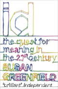
ID
by Susan GreenfieldIf you’ve ever wondered what effect video games have on your children’s minds or worried about how much private information the government and big companies know about you, ID is essential reading.Professor Susan Greenfield argues persuasively that our individuality is under the microscope as never before; now more then ever we urgently need to look at what we want for ourselves as individuals and for our future society.ID is an exploration of what it means to be human in a world of rapid change, a passionately argued wake-up call and an inspiring challenge to embrace creativity and forge our own identities.
ID
by Susan GreenfieldIf you?ve ever wondered what effect video games have on your children?s minds or worried about how much private information the government and big companies know about you, ID is essential reading.Professor Susan Greenfield argues persuasively that our individuality is under the microscope as never before; now more then ever we urgently need to look at what we want for ourselves as individuals and for our future society.ID is an exploration of what it means to be human in a world of rapid change, a passionately argued wake-up call and an inspiring challenge to embrace creativity and forge our own identities.
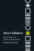
Idea Colliders: The Future of Science Museums (metaLAB Projects)
by Michael John GormanA provocative call for the transformation of science museums into "idea colliders" that spark creative collaborations and connections.Today's science museums descend from the Kunst-und Wunderkammern of the Renaissance--collectors' private cabinets of curiosities--through the Crystal Palace exhibition of 1851 to today's "interactive" exhibits promising educational fun. In this book, Michael John Gorman issues a provocative call for the transformation of science museums and science centers from institutions dedicated to the transmission of cultural capital to dynamic "idea colliders" that spark creative collaborations and connections. This new kind of science museum would not stage structured tableaux of science facts but would draw scientists into conversation with artists, designers, policymakers, and the public. Rather than insulating visitors from each other with apps and audio guides, the science museum would consider each visitor a resource, bringing questions, ideas, and experiences from a unique perspective.

The Idea Machine: My Inventor's Journal
by Max Grossman Jeffrey B. FuerstMax is a boy inventor searching for his next bright idea. He keeps a journal to help him find his eureka moment. Read Max's journal to find out what he comes up with.
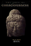
Idea of Consciousness: Synapses and the Mind
by Max R. BennettThe Idea of Consciousness examines the problem of how the working of synaptic connections might give rise to consciousness, and describes the current neuroscientific concepts and techniques used to identify and explore those parts of the brain that may be involved. This book will serve as an invaluable and stimulating introduction to the subject. Beautifully illustrated, it is a must for anyone who is curious about consciousness.
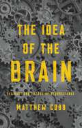
The Idea of the Brain: The Past and Future of Neuroscience
by Matthew CobbA powerful examination of what we think we know about the brain and why -- despite technological advances -- the workings of our most essential organ remain a mystery. For thousands of years, thinkers and scientists have tried to understand what the brain does. Yet, despite the astonishing discoveries of science, we still have only the vaguest idea of how the brain works. In The Idea of the Brain, scientist and historian Matthew Cobb traces how our conception of the brain has evolved over the centuries. Although it might seem to be a story of ever-increasing knowledge of biology, Cobb shows how our ideas about the brain have been shaped by each era's most significant technologies. Today we might think the brain is like a supercomputer. In the past, it has been compared to a telegraph, a telephone exchange, or some kind of hydraulic system. What will we think the brain is like tomorrow, when new technology arises? The result is an essential read for anyone interested in the complex processes that drive science and the forces that have shaped our marvelous brains.
The Idea of the Brain: A History
by Matthew CobbThis is the story of our quest to understand the most mysterious object in the universe. Today we tend to picture the brain as a computer. Earlier scientists thought about it in their own technological terms: as a telephone switchboard, or a clock, or all manner of fantastic mechanical or hydraulic devices. Could the right metaphor unlock the brain's deepest secrets once and for all?Galloping through centuries of wild speculation and ingenious, sometimes macabre anatomical investigations, scientist and historian Matthew Cobb reveals how we came to our present state of knowledge. Our latest theories allow us to create artificial memories in the brain of a mouse, and to build AI programmes capable of extraordinary cognitive feats. A complete understanding seems within our grasp.But to make that final breakthrough, we may need a radical new approach. At every step of our quest, Cobbshows that it was new ideas that brought illumination. Where, he asks, might the next one come from? What will it be?
The Idea of the Sciences in the French Enlightenment: A Reinterpretation (G - Reference, Information And Interdisciplinary Subjects Ser.)
by G. Matthew AdkinsThis book traces the development of the idea that the sciences were morally enlightening through an intellectual history of the secrétaires perpétuels of the French Royal Academy of Sciences and their associates from the mid-seventeenth century to the end of the eighteenth century. Academy secretaries such as Fontenelle and Condorcet were critical to the emergence of a central feature of the narrative of Enlightenment in that they encouraged the notion that the “philosophical spirit” of the Scientific Revolution, already present among the educated classes, should guide the necessary reformation of society and government according to the ideals of scientific reasoning. The Idea of the Sciences also tells an intellectual history of political radicalization, explaining especially how the marquis de Condorcet came to believe that the sciences could play central a role in guiding the outcome of the Revolution of 1789. Published by University of Delaware Press. Distributed worldwide by Rutgers University Press.
The Idea of War and Peace: The Experience of Western Civilization (Comparative Policy Evaluation Ser.)
by Irving HorowitzModern theorists and their ideas on war and peace are here presented, interpreted, and evaluated with scholarship and clarity of expression. In examining the main currents in modern social theory, the author has gone directly to the works of the leading philosophic figures. This book is a carefully documented analysis based on primary sources. Its republication in an expanded version after more than a half century since its initial appearance is a welcome addition to the literature on conflict and conflict resolution.In this 2007 greatly expanded third edition of The Idea of War and Peace, Irving Louis Horowitz provides a sense of substance to the character of Western Civilization. The book permits the reader to better understand what the "clash of civilizations" is about. It provides a broad outline of both European and American twentieth century social philosophies as they relate to the issue of war and peace. It also offers a new concluding section that explores in depth this same theme in the first decade of the twenty-first century.Such major figures as Bertrand Russell, John Dewey, Jacques Maritain, Albert Einstein, and Vladimir Lenin, reviewed in earlier editions, are now joined by examinations of the work of Raymond Aron, Harold D. Lasswell, and other contemporaries. The Idea of War and Peace is not just one more manual of how to conduct or avoid conflict, and even less, a guideline to policy-making. Instead, the work offers a profound sense of the theories and values that underline manuals and guides.This third edition is graced by a consideration of major figures in the second half of the twentieth century and a retrospective on the work of Niccolo Machiavelli on the nature of warfare. It also includes chapters on the relationship of war, peace, and the democratic order--and a postscript on new forms of state power and terrorism. This new edition links past and present and serves as an analytical bridge between cen
Ideal MHD
by Jeffrey FreidbergComprehensive, self-contained, and clearly written, this successor to Ideal Magnetohydrodynamics (1987) describes the macroscopic equilibrium and stability of high temperature plasmas - the basic fuel for the development of fusion power. Now fully updated, this book discusses the underlying physical assumptions for three basic MHD models: ideal, kinetic, and double-adiabatic MHD. Included are detailed analyses of MHD equilibrium and stability, with a particular focus on three key configurations at the cutting-edge of fusion research: the tokamak, stellarator, and reversed field pinch. Other new topics include continuum damping, MHD stability comparison theorems, neoclassical transport in stellarators, and how quasi-omnigeneity, quasi-symmetry, and quasi-isodynamic constraints impact the design of optimized stellarators. Including full derivations of almost every important result, in-depth physical explanations throughout, and a large number of problem sets to help master the material, this is an exceptional resource for graduate students and researchers in plasma and fusion physics.
The Ideal of Nature: Debates about Biotechnology and the Environment
by Gregory E. KaebnickIn this provocative anthology, scholars consider the meaning and merits of “nature” in debates about biotechnology and the environment.Drawing on philosophy, religion, and political science, this book asks what the term “nature” means, how it should be considered, and if it is—even in part—a social construct. The contributors question if the quality of being “natural” is intrinsically valuable. They also discuss whether appeals to nature can and should affect public policy and, if so, whether they are moral trump cards or should instead be weighed against other concerns.Though consensus on these questions remains elusive, this should not be an obstacle to moving the debate forward. By bringing together disparate approaches to addressing these concepts, The Ideal of Nature suggests the possibility of intermediate positions that move beyond the usual full-throated defense and blanket dismissal found in much of the debate. Scholars of bioethics, environmental philosophy, religious studies, sociology, public policy, and political theory will find much merit in this book’s lively discussion.