- Table View
- List View
Atlas of Laparoscopic Gynecological Anatomy
by Helizabet Salomão Ayroza Paulo Ayroza RibeiroThis book allows readers to gain a comprehensive understanding of gynecological surgical anatomy from a laparoscopic perspective. In recent years, with the growing number of gynecological surgical procedures performed by laparoscopy, many surgeons are faced with a “new” anatomy, not yet presented in traditional books. Addressing this gap in the literature and written in a colloquial style, this book presents much-needed information, especially regarding the spaces and the vessels, as well as numerous surgical tips and tricks. Focusing on retroperitoneal dissection, gynecological oncology and endometriosis, the book is intended for surgeons (gynecologists, urologists, general surgeons and others) interested in performing advanced pelvic surgery, offering them insights into how to transfer their knowledge of the traditional open surgery anatomy to the laparoscopic anatomy. Further, the book addresses 2D visualization and changes in the angle of visualization. The Atlas of Laparoscopic Gynecological Anatomy includes photos, surgical videos, drawings and figures to help readers quickly grasp the new concepts and to enhance the teaching power of the text.
Atlas of Laparoscopic Urologic Surgery
by Jan Roigas Serder Deger Stefan A Loening Andreas H WilleIdeal not only for the beginners in the field, but also for urologists looking to expand their knowledge, this clearly presented book adopts a useful step-by-step format to present established laparoscopic procedures. Easy and quick to use, the text includes a wealth of illustrations with important instructions, as well as tips and tricks described
Atlas of Laparoscopic and Robotic Single Site Surgery (Current Clinical Urology)
by Robert J. Stein Jihad H. Kaouk Georges-Pascal HaberThis text provides a broad and current review of this field and will serve as a valuable resource for trainees, academic and community surgeons, and members of industry with an interest in LESS. Due to the novelty and complexity of these procedures, the book focuses on detailed descriptions as well as pertinent illustrations for various upper and lower tract urologic procedures. The development of novel minimally invasive and robotic technology for more comfortable performance of these demanding procedures is covered. A complete description of instrumentation, platforms, and optics developed specifically for LESS is another primary focus of this text. Finally, a description of outcomes and complications as well as comparative data defining the status of LESS in relation to other current minimally invasive techniques is offered. Atlas of Laparoscopic and Robotic Single Site Surgery will provide a detailed summary of the current status of LESS that will help guide surgical decision making, encourage investigative efforts, and stimulate industry led technology development.
Atlas of Laparoscopy and Hysteroscopy Techniques
by Togas TulandiThis beautifully illustrated book provides a practical step-by-step guide to all the laparoscopic and hysteroscopic procedures performed by gynecologists. Each procedure is described in detail and fully illustrated with color photographs. The potential complications are described, and the circumstances in which a procedure is contraindicated are ex
Atlas of Lasers and Lights in Dermatology
by Keyvan Nouri Giovanni Cannarozzo Steven Paul Nisticò Mario SanninoThis richly illustrated atlas written by a team of experts guides the reader to the applications of lasers and light technologies in dermatology. It is divided in two parts: the first reviews the physical and optical concepts related to lasers and light sources, and provides a detailed description of surgical (ablative and non-ablative), vascular and pigmentary laser devices. It also discusses difficult-to-treat conditions, such as melasma and scars. The second part of the atlas is more clinically-oriented, presenting reproducible parameters and high-resolution images of pre and post-treatment, and desired end points in order to achieve an optimal result. Enabling readers to gain an understanding of the various topics concerning lasers, it explores conventional, non-conventional and combined laser treatments in a wide range of indications, as well as practical aspects such as medicolegal issues, informed consent and management of complications. The increasing knowledge and growing expertise in lasers and light devices make it necessary for physicians to be aware of the latest developments in this quickly evolving field. As such, this book is of interest to all physicians working in dermatology, cosmetology and aesthetic medicine, as well as to physician assistants and nurses using lasers in their daily practice.
Atlas of Leprosy
by Shekhar Neema Biju Vasudevan Santoshdev P Rathod Senkadhir Vendhan Akshay AmbasanaAtlas of Leprosy comprehensively covers the varied manifestations, including common and uncommon presentations, of leprosy, supported by patient photographs and additional video content, to assist medical practitioners diagnose leprosy in the early stages.With a detailed guide to approaching bedside diagnostic tests, this book will help readers to visualise common and uncommon presentations of leprosy, enabling effective diagnosis and treatment in this pre-elimination era, where increasingly difficulties in identifying its early stages can lead to preventable morbidity and deformity.Clinicians, dermatologists and trainees alike will find the wealth of high-quality illustrations and video demonstrations a vital guide to better diagnose this mycobacterial disease and combat resistance, relapse, reactions and any other difficult challenges posed.
Atlas of Limb Deformity: Etiological Classification
by Sihe Qin Wen Tian Qianyu Zhuang Baofeng GuoThis book focuses on classification and category of etiology, clinical manifestations, diagnosis, and orthopedic principles of different types of limb deformities in China. The relativity of the definition of limb deformity, the cognitive history of human beings on limb deformity and disability for thousands of years, and the characteristics and classification methods of the diseases category of limb deformity in Chinese population are covered in the chapters, in which the dynamic nature of the occurrence and development of limb deformity from the perspective of biological evolution and auxology, is analyzed too. Written by experts with wealthy of experience, it will be an ideal reference for physicians involved in the diagnosis and treatment of limb deformity.
Atlas of Lip and Nose Plastic and Cosmetic Surgery
by Jianhua Liu Bing ShiThis Atlas covers various types of lip and nose deformities and introduces individualized surgical plan for patients. The surgical techniques are classified according to the various types of deformities. Schematic diagrams, pre-operative and post-operative photographs are supplemented by detailed explanations. It consists of four parts: anatomical structure of lips and nose; cosmetic surgery of lips and nose; plastic surgery of nasolabial congenital deformity; plastic surgery of acquired nasolabial defect and deformities.The management of nasolabial deformities and defects crosses many surgical disciplines, and therefore, this book recommended to be read by surgeons of different backgrounds i.e. oral and maxillofacial surgery, plastic surgery, cosmetic surgery, otorhinolaryngology, etc. It can also be used as a reference for anatomy, developmental biology, physiology, linguistics, aesthetics and other basic medical sciences.
Atlas of Liquid Biopsy: Circulating Tumor Cells and Other Rare Cells in Cancer Patients’ Blood
by Ludmilla Thomé Domingos ChinenThis book provides a wealth of images and extensive information on circulating tumor cells (CTCs) and other cells usually observed in blood from patients with cancer, such as giant macrophages, and circulating tumor microemboli (CTM). The field of “liquid biopsy” began with the analysis of CTCs in the early 2000s. In the beginning, molecular techniques were developed to detect these cells in the blood. However, it has since become clear that CTCs initially require a cytopathological analysis to be detected without false positive and negative results. After detection, molecular analysis can be subsequently performed. Cancer is an important cause of mortality, especially when detected in late stages. Even with all the advances that have been made in its treatment, cancer is still challenging, as many patients do not respond to any therapy. Many health agencies have considered early diagnosis as a feasible tool. In this context, it is of the utmost importance to know the morphology and characteristics of CTCs to determine a correct diagnosis. Currently much of the scientific community is committed to expanding our knowledge of CTCs, and this work makes a valuable contribution, presenting hundreds of cell images from patients with various types of cancer, in many different stages of disease, and after receiving various treatments. The Atlas of Liquid Biopsy: Circulating Tumor Cells and Other Rare Cells in Cancer Patients’ Blood is an essential reference guide for all physicians, biologists, biomedics, and professionals working at clinical and research laboratories, hospitals and research centers.
Atlas of Liver Pathology (Atlas of Anatomic Pathology)
by Anthony W. H. Chan Alberto Quaglia Beate Haugk Alastair BurtThe liver is a complex organ due to its unique microscopic structure, intricate metabolic functions and susceptibility to a wide variety of insults, manifesting in countless histological patterns. Atlas of Liver Pathology considers both changes seen in medical liver biopsies as well as lesional biopsies when the specimen has been taken from a mass. The book starts by reviewing normal structure and its variants and the optimal approaches for the preparation of histological sections for diagnostic liver pathology. The following chapters are dedicated to developmental, metabolic, infectious, drug related, autoimmune, biliary, vascular and neoplastic disorders. Two sections on liver pathology in pregnancy and transplantation conclude the work. Macroscopic illustrations are included where appropriate. All photographs are complemented by legends describing the picture and providing relevant related information. Authored by nationally and internationally recognized pathologists, Atlas of Liver Pathology is a valuable resource that serves as a quick reference guide for the diagnosis of usual and unusual diseases.
Atlas of Liver Pathology: A Pattern Based Approach To Neoplastic Biopsies
by Michael TorbensonPart of the new Pattern-Based Approach series, designed to teach pathology in a way that reflects actual sign-out, this new visually-oriented guidebook presents real-life cases and practical diagnostic tips. Disease processes are organized into ‘patterns’ of injury—the method by which pathologists approach their work—and self-assessment quizzes are provided for each chapter to give you experience with high-yield, board-style teaching topics.
Atlas of Liver Transplantation
by Daniel Azoulay Chetana Lim Chady SalloumThe Atlas of Liver Transplantation presents the principles and techniques of liver transplantation in a vividly illustrated, easy-to-understand format. Its primary objective is to portray common situations in liver transplantation: from graft harvesting to explantation of the native liver, portocaval anastomosis to biliary anastomosis. Featuring more than 350 color photographs organized into thirteen chapters, Atlas of Liver Transplantation is designed to be equally useful for students, surgical residents, trainees, surgeons seeking current evidence-based information in subspecialty areas relevant to their general surgical practice, and leading experts in the field.

Atlas of Living Cell Cultures
by Rosemarie Steubing Toni LindlThe first atlas in many years giving researchers a good visual reference of the status of their cell lines. Given the increasing importance of well defined cellular models in particular in biomedical research this is a sorely needed resource for everyone performing cell culture.
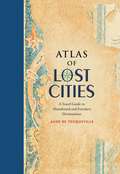
Atlas of Lost Cities: A Travel Guide to Abandoned and Forsaken Destinations
by Aude De TocquevilleLike humans, cities are mortal. They are born, they thrive, and they eventually die. In Atlas of Lost Cities, Aude de Tocqueville tells the compelling narrative of the rise and fall of such notable places as Pompeii, Teotihuacán, and Angkor. She also details the less well known places, including Centralia, an abandoned Pennsylvania town consumed by unquenchable underground fire; Nova Citas de Kilamba in Angola, where housing, schools, and stores were built for 500,000 people who never came; and Epecuen, a tourist town in Argentina that was swallowed up by water. Beautiful, original artwork shows the location of the lost cities and depicts how they looked when they thrived.
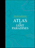
Atlas of Lost Paradises (Atlas Series #3)
by Gilles LapougeThoroughly documented, a worldwide selection of places representing many attempts made by mankind through the ages to re-create a paradise on Earth. "Paradises got off to a bad start early on. The one the Bible had arranged had to rapidly close its pearly gates when its first two occupants, Adam and Eve, had misbehaved." According to Gilles Lapouge, paradise is a paradoxical creation of our imagination, blending hope and nostalgia. Historically, mankind has sought to fashion a paradise, which could be accessed during its lifetime: ideal cities, cities made of glass and steel, castles of freedom, etc. This atlas embarks us upon a journey across civilizations, through the exploration of 27 real or fictional places, including • gardens of the Middle Ages • Atlantis • the castles of King Ludwig II • Oceana • Pitcairn Island • city of Manoa • Mausoleum of Qin Shi Huang Each place is illustrated with a specially designed map in a graphic style that has become the hallmark of this Atlas series.
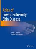
Atlas of Lower Extremity Skin Disease
by Tracey C. Vlahovic Stephen M. SchleicherLower extremity skin disorders are often overlooked by clinicians. Ailments such as eczema, psoriasis and tinea at times prove difficult to distinguish clinically, and misdiagnosis can lead to inappropriate therapy. Many practitioners are mystified when confronted with an abnormal appearing nail. Delay in recognizing skin cancer may adversely impact morbidity and mortality. This full-color atlas is a concise guide for medical professionals who deal with the lower extremities and will aid in both diagnosis and treatment. Topics featured in the Atlas include nail pathology, fungal and bacterial infections, xerotic and hyperkeratotic disorders, autoimmune diseases and vasculopathies, benign and malignant lesions, systemic diseases, and ulcerations. Each chapter contains vibrant photographic representative examples. Concluding chapters present a review of biopsy techniques as well as an overview of current dermatological therapies. The Atlas of Lower Extremity Skin Disease is a unique resource for podiatrists, dermatologists, and primary care physicians as well as nurse practitioners and physician assistants.
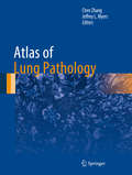
Atlas of Lung Pathology (Atlas of Anatomic Pathology)
by Chen Zhang Jeffrey L. MyersAtlas of Lung Pathology offers a comprehensive survey of common and uncommon conditions likely to be encountered in a wide range of practices. It begins with a review of normal macroscopic and microscopic lung anatomy followed by practical tips on how to handle the most common specimens received in pathology practice. Non-neoplastic conditions are segregated into logical categories intended to make them easily accessible to quickly offer solutions for diagnostic problems encountered in daily practice. Each illustration was carefully chosen using images drawn from experienced contributors familiar with the challenges of diagnostic pulmonary pathology.
Atlas of Lymph Node Anatomy
by Mukesh G. HarisinghaniDetailed anatomic drawings and state-of-the-art radiologic images combine to produce this essential Atlas of Lymph Node Anatomy. Utilizing the most recent advances in medical imaging, this book illustrates the nodal drainage stations in the head and neck, chest, and abdomen and pelvis. Also featured are clinical cases depicting drainage pathways for common malignancies. 2-D and 3-D maps offer color-coordinated representations of the lymph nodes in correlation with the anatomic illustrations. This simple, straightforward approach makes this book a perfect daily resource for a wide spectrum of specialties and physicians at all levels who are looking to gain a better understanding of lymph node anatomy and drainage. Edited by Mukesh G. Harisinghani, MD, with chapter contributions from staff members of the Department of Radiology at Massachusetts General Hospital.
Atlas of Lymph Node Anatomy
by Mukesh G. HarisinghaniThis book is a comprehensive atlas on lymph node anatomy and drainage to aid in cancer staging and therapy. Nodal drainage is pertinent to all aspects of cancer staging and therapy and is used by radiation oncologists, surgeons, and medical oncologists to increase accuracy. The first edition of this text was the first comprehensive monograph on this topic, allowing physicians across various specialties to utilize this information and easily share that knowledge with residents, fellows, and junior faculty.Detailed anatomic drawings and state-of-the-art radiologic images combine to produce this essential Atlas of Lymph Node Anatomy. Utilizing the most recent advances in medical imaging, this book illustrates the nodal drainage stations in the head and neck, chest, abdomen, and pelvis. Also featured are clinical cases depicting drainage pathways for common malignancies. 2-D and 3-D maps offer color-coordinated representations of the lymph nodes in correlation with the anatomic illustrations. This simple, straightforward approach makes this book a perfect daily resource for a wide spectrum of specialties and physicians at all levels who are looking to gain a better understanding of lymph node anatomy and drainage.This new edition enables physicians to educate themselves on the location of various nodal stations, especially in the context of common primary tumors, so that they are able to detect, localize, and characterize nodes seen with novel new imaging methods and with an increased level of accuracy. Chapters now cover the significant strides made in the imaging realm, such as PET CT, conventional MRI, MRI with novel imaging agents, and multidetector CT, which allows visualization of lymph nodes in various anatomic compartments.
Atlas of Lymph Node Pathology (Atlas of Anatomic Pathology)
by Joseph D. Khoury L. Jeffrey Medeiros Roberto N. MirandaAtlas of Lymph Node Pathology reviews the histopathology of nodal diseases, illustrating the use of ancillary studies and includes concise discussions of pathogenesis, clinical settings and clinical significance of the pathologic diagnosis. The atlas features an overview of the benign reactive processes secondary to infectious, environmental or unknown insults, as well as relevant illustrations of virtually all primary and secondary neoplasms involving lymph nodes. The atlas also includes macroscopic images of some disorders, tables that help readers understand and comprehend diseases that look alike, and diagnostic algorithms for certain groups of diseases. Authored by highly experienced pathologists, Atlas of Lymph Node Pathology is a valuable resource that illustrates the vast majority of diseases practicing pathologists, clinicians and oncologists are likely to encounter in daily practice.
Atlas of Lymph Node Pathology: A Pattern Based Approach
by Amy S. Duffield Joo Y. Song Girish VenkataramanClosely mirroring the daily sign-out process, Atlas of Lymph Node Pathology: A Pattern Based Approach is a highly illustrated, efficient guide to accurate diagnosis. This practical reference uses a proven, pattern-based approach to clearly explain how to interpret challenging cases by highlighting red flags in the clinical chart and locating hidden clues in the slides. Useful as a daily “scope-side guide,” it features numerous clinical and educational features that help you find pertinent information, reach a correct diagnosis, and assemble a thorough and streamlined pathology report.
Atlas of Lymphatic Anatomy in the Head, Neck, Chest and Limbs
by Wei-Ren PanThis atlas provides detailed information on the human lymphatic system in the head, neck and chest regions as well as the extremities, with more than 400 photographs and radiographs, including micro and macro views of the morphology. Much of the content is presented for the first time, such as the individual differences in lymphatic distribution, especially in the head neck region; characteristics of the indirect precollecting lymph vessel in the scalp; the lymphatic ampulla and diverticulum; and the transparent lymph node. Providing insights into the lymphatic anatomy, the book is an essential resource for medical and science students as well as therapists, clinicians and researchers working in this field.
Atlas of Lymphatic System in Cancer: Sentinel Lymph Node, Lymphangiogenesis and Neolymphogenesis
by Shamil Gantsev Kamil Gantsev Shamil KzyrgalinThis atlas contains a large number of original photographs illustrating the anatomy, research methods, and structural features of the structure of the lymphatic apparatus in oncological diseases. The tissue complexes of the axillary region have been studied in detail. The lymphatic maps of the axillary region in cancer are presented, including those with manifestations of lymphangiogenesis; newly formed lymph nodes formed as a result of neolymphogenesis are also considered. Atlas of Lymphatic System in Cancer: Sentinel Lymph Node, Lymphangiogenesis and Neolymphogenesis is intended for oncologists, anatomists, morphologists and doctors of other specialties. The book is the result of ten years of research. The applied technology of ultrasonic isolation of anatomical structures made it possible to take a fresh look at the lymphatic apparatus in cancer and describe the phenomenon of neolymphogenesis.
Atlas of Lymphoscintigraphy and Sentinel Node Mapping: A Pictorial Case-Based Approach
by Giuliano Mariani Sergi Vidal-Sicart Renato A. Valdés OlmosThe main goal of the second edition of this book is to update the content on the rapidly growing field of lymphoscintigraphy, a radionuclide-based imaging procedure that provides information on the functional status of the lymphatic system. Although the technique was originally introduced to identify the cause of peripheral edema (i.e., blockage of the venous or lymphatic circulation), more recent and widespread applications include radioguided biopsy of the sentinel lymph node in patients with solid cancers. This procedure is crucial for the adequate planning of oncologic surgery in a growing number of cancers, most notably breast cancer, cutaneous melanoma, head and neck cancers, penile cancer, and cervical cancer.The book focuses on the latest advances in lymphoscintigraphy techniques, including both novel tracers recently approved for clinical use (especially in the field of sentinel lymph node mapping) and the expanding role of hybrid imaging with SPECT/CT – and in sentinel node detection using hybrid tracers (radiolabeled and fluorescent) for dual-signature guidance. Each chapter addresses the clinical application of lymphoscintigraphy in different anatomic areas or disease conditions. After an introductory section concerning the pathophysiology of the specific site/disease, the clinical relevance and impact of lymphoscintigraphy is demonstrated by a collection of richly illustrated teaching cases describing the lymphoscintigraphic patterns most commonly observed, as well as anatomic variants and technical pitfalls. Emphasis is placed on tomographic multimodality imaging. The book gathers contributions by experts in nuclear oncology, who have revised their chapters by updating the didactic material and adding clinical cases. Regarding sentinel lymph node biopsy in particular, a major distinction of this text is the incorporation of the staging guidelines of the American Joint Committee on Cancer (8th edition) into the didactic material.
Atlas of Macroscopic Wood Identification: With a Special Focus on Timbers Used in Europe and CITES-listed Species
by Alan Crivellaro Flavio RuffinattoThis atlas presents macroscopic descriptions, macro cross section pictures, general characteristics and identification keys of 335 wood species currently introduced in the European timber market from all over the world. Overall 292 different genera are represented and CITES-listed timbers are also included. Macroscopic descriptions are based on a recently proposed list of macroscopic features for wood identification. Macroscopic features and their codes are defined and illustrated in the atlas. Wood descriptions also include information about natural durability, physical and mechanical properties, end uses, environmental sustainability and possible related misleading commercial names. Furthermore, each genus is described in terms of number of species, geographical distribution and main commercial timbers, and details are given about to what extent timbers within the genus can be typically identified through macroscopic and microscopic analysis, if any.The atlas will be a valuable guide for all agents in charge for timber verification, those involved in the European Timber Regulation enforcement and CITES inspections, as well as wood scientists, foresters, wood sellers, wood restorers, and any wood worker and wood passionate interested in a fast and reliable tool for wood identification.