- Table View
- List View
Atlas of Operative Procedures in Surgical Oncology
by Constantine P. KarakousisThis volume is the product of the author's long experience with melanomas and sarcomas and to a lesser but significant degree with upper gastrointestinal cancers, colorectal and breast cancers. As such, it offers a "hands-on" practical guide to approaching complex soft-tissue tumors and for performing more extensive tumor resections based on over 40 years of surgical experience. It provides important details about the positioning of patients, incision types, and exposure which can be of paramount importance in the resection of certain tumors. The book contains important general surgical principles for approaching tumors in a variety of locations but also offers the detail necessary for the safe and oncologically sound resection of these malignancies. Moreover, this operative atlas contains specific information for procedures which are not as commonly encountered in surgical training, but can be invaluable in the management of patients with locally aggressive tumors, such as hemipelvectomy and its variants, sacral resections, and forequarter amputation. Through the use of multiple detailed illustrations, Atlas of Operative Procedures in Surgical Oncology serves as a valuable resource to the general surgeon or surgical oncologist in the operative management of patients with cancer in the abdomen, retroperitoneum, pelvis or extremities.
Atlas of Operative Techniques in Primary Cleft Lip and Palate Repair
by Percy Rossell-PerryFew books have been published before with details on preoperative planning, markings and performance of primary surgical techniques to correct the cleft lip and palate deformity. This information is commonly required by surgeons. Scientific papers, conferences, and video clips of these surgical procedures are limited in details about how to address this common disease.This book provides a comprehensive overview of surgery for the correction of primary cleft lip and palate, including planning, selecting the most suitable techniques, markings, performing operative techniques, and preoperative care. Unlike other literature on the topic, which focuses on surgical techniques, this operative atlas (with detailed illustrations) covers the entire spectrum of this congenital deformity: classification of cleft lip and palate, management protocols, unilateral cleft lip surgery, bilateral cleft lip surgery, cleft palate surgery, post operative care and case studies. It will be a unique and valuable resource for surgeons treating this common condition.
Atlas of Optical Coherence Tomography for Glaucoma
by Donald L. BudenzAtlas of Optical Coherence Tomography for Glaucoma is a case-based atlas intended to teach the reader how to interpret the results of OCT in glaucoma patients and glaucoma suspects. After a brief description of how OCT is used in particular situations, chapters depict actual case presentations from authors’ practices with legends that describe the case and how OCT is used to make the diagnosis of glaucoma or glaucoma progression. Emphasis is placed on where OCT can lead the clinician astray by providing false positive or false negative results resulting in misdiagnosis. The intention of the format is to make it easily digestible in a weekend read and make the practitioner comfortable with OCT interpretation. Examples are presented from all of the available OCT manufacturers.
Atlas of Oral Diseases: A Guide for Daily Practice
by Isaäc van der WaalThis atlas is designed to assist all who are involved in diagnosing and treating oral diseases. Individual chapters focus on lesions and disorders of the oral mucosa, soft tissues (including the minor salivary glands), lips, tongue, gingiva, palate, and jaw bones (odontogenic and non-odontogenic lesions). In addition to the more common diseases, less frequent disorders are also covered, some of which have been recognized only in recent years. Throughout, the approach is practice oriented, with concise text and an abundance of high-quality clinical, radiographic, and, where appropriate, histopathologic images. The combined training of the author in oral surgery and oral pathology means that he has exceptional expertise in both the diagnosis and the treatment of oral diseases. His detailed knowledge and experience are fully reflected in the Atlas of Oral Diseases, which will be very helpful for dental and medical professionals in their daily practice.
Atlas of Oral Microbiology: From Healthy Microflora To Disease
by Xuedong Zhou Yuqing LiThis book is the second edition of Atlas of Oral Microbiology: From Healthy Microflora to Disease (ISBN 978-0-12-802234-4), with two new features: we add about 60 pictures of 14 newly isolated microbes from human dental plaque, at the same time, we re-organize the content of this book and provide more research progress about the oral microbiome bank of China, the invasion of oral microbiota into the gut, and the relationships between Oral Microflora and Human Diseases. This book is keeping up with the advanced edge of the international research field of oral microbiology. It innovatively gives us a complete description of the oral microbial systems according to different oral ecosystems. It collects a large number of oral microbial pictures, including cultural pictures, colonies photos, and electron microscopy photos. It is by far the most abundant oral microbiology atlas consists of the largest number of pictures. In the meantime, it also described in detail a variety of experimental techniques, including microbiological isolation, culture, and identification. It is an atlas with strong practical function. The editors and writers of this book have long been engaged in teaching and research work in oral microbiology and oral microecology. This book deserves a broad audience, and it will meet the needs of researchers, clinicians, teachers, and students major in biology, dental medicine, basic medicine, or clinical medicine. It can also be used to facilitate teaching and international academic exchanges.
Atlas of Oral and Maxillofacial Anatomy
by R. Shane Tubbs Joe IwanagaThis comprehensive atlas, featuring a wealth of top quality photographs of fresh cadaveric dissections, is a superb guide to anatomic structures in the oral and maxillofacial region that will be an ideal aid in clinical practice. It has the important benefit of enabling readers to observe the anatomy from the same view as seen during invasive clinical procedures. This is critical for a better understanding of these procedures, and surgical annotations are included as necessary. Atlas of Oral and Maxillofacial Anatomy is the first book of its kind to be devoted to the clinical anatomy of the region for dentists and oral and maxillofacial surgeons. It will satisfy the demand for such a comprehensive atlas in this field of surgery and will be welcome and timely for clinicians and trainees. Beyond specialists and residents in oral and maxillofacial surgery and general dentists, the book will be of value for craniofacial surgeons, anatomists, plastic surgeons, ENT surgeons, head and neck surgeons, neurosurgeons, dental students, medical students, dental hygienists, and nurses working with dentists and oral and maxillofacial surgeons.
Atlas of Oral and Maxillofacial Radiology
by Bernard KoongThe Atlas of Oral and Maxillofacial Radiology presents an extensive case collection of both common and less common conditions of the jaws and teeth. Focusing on the essentials of radiologic interpretation, this is a go-to companion for clinicians in everyday practice who have radiologically identified a potential abnormality, as well as a comprehensive study guide for students at all levels of dentistry, surgery and radiology. Unique lesion-based problem solving chapter makes this an easy-to-use reference in a clinical setting Includes 2D intraoral radiography, the panoramic radiograph, cone beam CT, multidetector CT and MRI Multiple cases are presented in order to demonstrate the variation in the radiological appearances of conditions affecting the jaws and teeth Special focus on conditions where diagnostic imaging may substantially contribute to diagnosis
Atlas of Orbital Imaging
by Guy Ben Simon Gahl Greenberg Daphna Landau PratThis book features in-depth descriptions of imaging modalities (MRI, CT, US, PET) for all orbital pathologies, including tumors, vascular anomalies, congenital anomalies, trauma, inflammations and infections. It describes all the imaging features of the pathologies, and includes guidance for differential diagnosis and relevant clinical data. Atlas of Orbital Imaging serves as a clinical and educational resource for ophthalmologists/orbital surgeon residents, as well as a source of reference for consultants and neuroradiologists at all levels. The illustrations are both highly detailed and depict the orbit in vivid colour, adding to the attractiveness of the chapters. This reference work is a worldwide collaborative effort of all leading orbital surgeons and neuro-radiologists (in Europe, America, Australia and Asia) and provides an indispensable resource for developing skills and knowledge of orbital imaging.
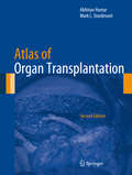
Atlas of Organ Transplantation
by Abhinav Humar Mark L. SturdevantAtlas of Organ Transplantation, Second Edition, provides the reader with a comprehensive and pictorial step-by-step account of abdominal organ transplant procedures performed by contemporary transplant surgeons today. Emphasis is placed on newer procedures or procedures that have undergone significant modifications. It is recognized that there are many well-accepted techniques for the same procedure, with each having potential merit. While it is impossible to present all of these variations, an attempt is made to describe the common variations in surgical technique and common variation in procedure based on anatomical variations. Written by an expert in the field, Atlas of Organ Transplantation, Second Edition, includes schematic diagrams and high-quality intraoperative photographs, allowing readers to clearly visualize the course of the operative procedure. This format provides the reader with a clear visual and written description of all major transplant procedures in one reference book.
Atlas of Orthodontic Case Reviews
by Marjan Askari Stanley A. AlexanderAtlas of Orthodontic Case Reviews offers a comprehensive resource to the treatment of orthodontic malocclusions with a case-based approach. Discusses and illustrates the treatment of orthodontic malocclusions using actual clinical cases Presents more than 800 clinical photographs showing the stages of each treatment, to act as a visual reference Includes a description of each malocclusion, an explanation of the desired treatment outcomes, an account of the changes, and review questions for each case
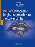
Atlas of Orthopaedic Surgical Approaches to the Lower Limbs
by Rosa Ballis Bujar H. Shabani Dafina BytyqiEnriched by original anatomical artworks this atlas comprehensively presents the diverse orthopaedic approaches to lower limb surgery. Combining clear visual information with a consistent and concise structure, including tips, tricks and pitfalls, this manual presents all the most used approaches in orthopaedic prosthetic surgery and traumatology. Ranging from the hip to the ankle each chapter includes a clear presentation of the joint’ s anatomy, with particular emphasis on vessels, nerves and other relevant anatomic structures. Beautiful water-colour illustrations realized by one of the authors accompany the reader step-by-step through each of the approaches described. This atlas offers a timely and up-to-date resource for both practicing and fellow orthopaedic surgeons with an interest in lower limb surgery.
Atlas of Osteopathic Techniques
by Alexander S. Nicholas Evan A. NicholasEasy to navigate and rich with engaging learning features, the 4th edition of this bestselling, one-of-a-kind resource reflects the most up-to-date information on basic anatomical concepts and techniques to help users confidently comprehend and apply them.
Atlas of PET-CT Imaging in Oncology: A Case-Based Guide to Image Interpretation
by Tamer Özülker Filiz ÖzülkerThis atlas is a case-based guide to the interpretation of FDG PET-CT images in clinical scenarios faced by physicians during the routine practice of oncology. The book aims to help the practitioner to overcome diagnostic dilemmas through familiarization with the physiologic distribution of FDG, normal variants and benign findings. The main focus, however, is the imaging of major oncological diseases. Different pathologies are addressed in individual chapters comprising teaching files of cases, each of which corresponds to a common indication for PET-CT imaging, such as metabolic characterization of lesions, staging, restaging and evaluation of response to therapy. Each case is accompanied by an explanation of the patient's history, interpretation of the PET-CT study, and a teaching point often supported by relevant literature. This book will be of great value to residents and practitioners in nuclear medicine, radiology, oncology, radiation oncology and nuclear medicine technology.
Atlas of PET/CT in Oncology - Volume 1: Brain, Head and Neck Cancers
by Zhiming Yao Sijin LiThe atlas aims to help practitioners to interpret PET/CT images of tumors in brain, head and neck in a timely and accurate way. Illustrating in a case-based manner, the PET/CT appearances of glioma, meningioma, nasopharyngeal carcinoma, eye tumors, ear and temporal bone tumors, and neck tumors are covered. Each chapter is organized in the same format of clinical overview, PET/CT diagnosis, typical/atypical/confusing manifestation of the diseases. This book will be a useful reference to residents and practitioners in nuclear medicine, radiology, oncology, radiation oncology and nuclear medicine technology.
Atlas of PET/CT in Pediatric Patients
by Angelina CistaroThis richly illustrated book presents the pediatric applications of PET/CT in the full range of scenarios frequently encountered in a professional setting. It opens with a thorough introduction covering the fundamental science and the clinical basis for use of PET/CT in this age group. Pitfalls and artifacts are examined, and normal variations and benign findings are carefully described. Each subsequent chapter addresses the role of PET/CT with different radiopharmaceuticals in the evaluation and management of a specific disease. The full range of oncological diseases is covered, including the rare ones. Succinct descriptions of clinical cases are included and, when appropriate, comparisons are made with other modalities. In addition, the role of PET/CT in biopsy guidance and in radiation therapy planning is explained. This book will be invaluable for residents and practitioners in nuclear medicine, radiology, oncology, radiation oncology, and nuclear medicine technology
Atlas of PET/MR Imaging in Oncology
by Osman Ratib Markus Schwaiger Thomas BeyerThis new project on PET-MR imaging in oncology includes digital interactive software matching the cases in the book. The interactive version of the atlas is based on the latest web standard, HTML5, ensuring compatibility with any computer operating system as well as a dedicated version for Apple iPad and iPhone. The book opens with an introduction to the principles of hybrid imaging that pays particular attention to PET/MR imaging and standard PET/MR acquisition protocols. A wide range of illustrated clinical case reports are then presented. Each case study includes a short clinical history, findings, and teaching points, followed by illustrations, legends, and comments. The multimedia version of the book includes dynamic movies that allow the reader to browse through series of rotating 3D images (MIP or volume rendered), display blending between PET and MR, and dynamic visualization of 3D image volumes. The movies can be played either continuously or sequentially for better exploration of sets of images. The editors of this state-of-the-art publication are key opinion leaders in the field of multimodality imaging. Professor Osman Ratib (Geneva) and Professor Markus Schwaiger (Munich) were the first in Europe to initiate the clinical adoption of PET/MR imaging. Professor Thomas Beyer (Zurich) is an internationally renowned pioneering physicist in the field of hybrid imaging. Individual clinical cases presented in this book are co-authored by leading international radiologists and nuclear physicians experts in the use of PET and MRI.
Atlas of Paediatric Surgical Imaging: A Clinical and Diagnostic Approach
by Robert CarachiThis book is a comprehensive compendium of paediatric conditions, and covers clinical and diagnostic imaging for most diseases affecting neonates and children. Detailed descriptions of radiological signs aim to aid the diagnosis and identification of clinical symptoms. The book contains a large number of images taken from a collection of current and archival photos obtained from three generations of paediatric surgeons and radiologists which further illustrate the points made in the text. This book will act as a reference manual for any person in training who has to care for neonates and children in a hospital setting.
Atlas of Palpation
by Robin Bauer Sandro WolframThis atlas with over 250 illustrations and videos is a modern basic work on palpation for physiotherapists. Members of the medical-therapeutic sector face the daily challenge of having to transfer theoretical knowledge into practice. The Atlas of Palpation addresses this process through its multimedia design. Evidence-based technical texts, the most modern illustrations and practical teaching videos illustrating structures and examination procedures address both students and therapists working in practice. All the essential structures of the body are shown in three-dimensional perspective, so that a basic understanding of interrelationships and movement patterns is conveyed. The palpation grips are explained in an understandable way and described in a comprehensible way. The instructional videos were specially made by the authors and the latest technical equipment in terms of camera, lighting elements and sound ensures the highest possible quality. The book is clearly divided into different body systems, which include bony, articular, ligamentous, muscular, nervous, and vascular system. A clear subdivision into subheadings makes it possible to quickly find the desired content. The multimedia approach of this book represents a unique selling point in the medical-therapeutic sector in the field of palpation. This new type of "living" book opens up completely new perspectives for readers when using it. Trainees and students will find here an optimal introduction for professional palpation; for already experienced physiotherapists it is an ideal reference book for tricky questions. Download the SN More Media app for free, scan a link with play button and access directly on your smartphone or tablet.
Atlas of Parathyroid Imaging and Pathology
by Alexander L. Shifrin L. Daniel Neistadt Pritinder K. ThindThis book provides a visual demonstration of normal and ectopic locations of parathyroid adenomas using different modalities in patients with PHPT and to describe parathyroid gland–related pathology. It includes several modern imaging modalities for localization of parathyroid glands and parathyroid adenomas, such as Sestamibi scan, SPECT/CT Sestamibi scan, neck ultrasound, MRI, thin-cut CT, and 4D CT scans. Written by experts in the field, chapters include pathology images corresponding to radiology imaging for some presented cases (gross and high-power view). Authors have also collected radiological images of difficult-to-localize parathyroid adenomas in ectopic (abnormal) locations. The atlas is organized by location of the adenomas in upper and lower eutopic locations followed by ectopic locations. Each case demonstrates dual or triple modalities such as US, Sestamibi scan, or SPECT/CT Sestamibi scan, thin-cut CT scan, or 4D CT performed on the same patient. A chapter on parathyroid pathology is also included to help the reader understand challenges in pathological interpretation. Atlas of Parathyroid Imaging and Pathology serves as a valuable reference for radiologists, endocrine surgeons, head and neck surgeons, ENT surgeons, surgical oncologists, endocrinologists, pathologists, nephrologists, students, and all physicians and allayed health practitioners involved in the treatment of patients with primary, secondary, and tertiary hyperparathyroidism.
Atlas of Parathyroid Surgery
by Alexander ShifrinThis Atlas is designed to illustrate different techniques on how to perform successful parathyroidectomy by using traditional four gland exploration approach and minimally invasive approaches, such as the open minimally invasive approach, video-assisted approach, back-door approach, transoral endoscopic parathyroidectomy approach (TOEPVA), and endoscopic lateral parathyroidectomy approach. It illustrates removal of a right and left, and superior and inferior parathyroid glands. Written by renowned endocrine surgeons and experts in the field, each chapter begins with a case description that defines the main aspect of surgery. Each picture, which is taken intraoperatively, is accompanied by corresponding drawings for easier understanding of the anatomical structures and steps of the procedure. In addition, most of the authors provided a video of the same case as it is depicted in the chapter. The Atlas also gives some common pitfalls of the procedure in an effort to avoid complications and improve patient outcomes. Atlas of Parathyroid Surgery provides an indispensable source of knowledge to all surgeons, those who just started their career, and those who are in the more advanced stages of their practice and are learning new techniques of parathyroidectomy.
Atlas of Pathologic Myopia
by Kyoko Ohno-MatsuiThis Atlas provides many beautiful images obtained with state-of-the-art technologies, including optical coherence tomography (OCT), OCT angiography, fundus autofluorescence, and wide-field fundus imaging, as well as traditional images and fluorescein/ICG angiograms. Gathered at the world’s largest High Myopia Clinic, the images are based on the long-term follow-up data of more than 6,000 patients from Japan and abroad. Recent advances in imaging technologies have yielded many new observations and allowed us to detect new lesions, e.g. myopic traction maculopathy (or macular retinoschisis) and dome-shaped macula. An especially interesting aspect: the images obtained by ‘3D MRI of the eye’ and ‘ultra wide-field OCT’ to visualize staphylomas. These techniques were established by the editor’s group and make it possible to record the entire shapes of the eye, offering a scan width of up to 23 mm and scan depth of 5 mm. They have since been used to visualize posterior staphyloma, which was previously impossible to view because it spanned such a wide range of the eye. In addition, readers will learn what types of eye deformity occur in pathologic myopia and how they damage the macula/optic nerve. With this Atlas, readers will learn how to accurately diagnose each lesion of pathologic myopia, how eye deformity causes blinding complications, and how to identify patients with a poor prognosis. In short, it provides essential information that can’t be found elsewhere.
Atlas of Peculiar and Common Testicular and Paratesticular Tumors
by Manuel Nistal Pilar González-PeramatoThis book, conceived as an atlas with emphasis in diagnostic images, presents some tumors displaying expected clinical, histological or biological behavior but in most of them that differs from that usual morphology or clinical behaviour. About 132 tumors of the testicles and paratesticular structures have been selected, representing all the testicular and paratesticular tumor groups, including germinal, gonadal stromal, non-gonadal stromal, rete testis tumors, ovarian-type epithelial tumors, and epididymis, spermatic cord and testicular covers as well as metastatic tumors.The format with large number of images allows pathologists to identify the entities included at a glance. Each section corresponds to one case including a brief clinical history, a concise histological description with immunohistochemical techniques necessary to confirm the diagnosis. In addition, each section provides several sample images, with histological details of the appropriate morphological or characteristic immunoexpression of diagnostic markers and in the majority of cases gross or radiologic figures. Finally, each section ends with a comment on the problems of differential diagnosis that could arise. The book is intended for pathologists, urologists and oncologists.
Atlas of Pediatric Brain Tumors
by Tina Young Poussaint Tarik Tihan Adekunle M. Adesina Christine E. FullerRecent evidence indicates that the physiology and response to various drugs in the pediatric population differs from that of the adult and must be appreciated to be able to fully address the health needs of the pediatric population. Atlas of Pediatric Brain Tumors covers areas ranging from neuroimaging, the use of crush and touch preps during introperative consultation, classic histological features of brain tumors, tumor variants, and a miscellaneous group of challenging tumors. Chapters consist of essential diagnostic information and features highlighting recognized variants and their differential diagnoses. A section on molecular pathology and electron microscopy is also included for each tumor category; along with a list of classic reviews and innovative articles on each of the tumor entities as suggested reading at the end of each chapter. Created to fill a void in the practice of pediatric neuropathology, this practical and well-illustrated Atlas of Pediatric Brain Tumors represents a collection of interesting, common and unusual tumors for a diagnostic exercise by the reader.
Atlas of Pediatric Brain Tumors
by Tina Young Poussaint Tarik Tihan Adekunle M. Adesina Christine E. FullerThis text was created to fill a void in the practice of pediatric neuropathology. It is a practical and well-illustrated book representing a collection of interesting, common and unusual tumors for a diagnostic exercise by the reader. The wide reception of the first edition by the pathology community is testament to its relevance and utility in the pathologic diagnosis of pediatric brain tumors. This edition covers topics ranging from neuroimaging, the use of crush and touch preps during intraoperative consultation, classic histological features of pediatric brain tumors, tumor variants, and a miscellaneous group of challenging tumors. Chapters consist of essential diagnostic information and features highlighting recognized variants and their differential diagnoses. A section on molecular pathology and electron microscopy is also included for each tumor category, along with a list of classic reviews and innovative articles on each of the tumor entities as suggested reading at the end of each chapter. Atlas of Pediatric Brain Tumors, Second Edition represents the state of the art in pediatric neuropathology with easy utility beside the microscope.
Atlas of Pediatric CTA of Coronary Artery Anomalies
by Randy Ray RichardsonWhile most current texts on the coronary artery include only a single chapter on anomalies, this is the first of its kind to focus on the challenges involved with imaging coronary artery anomalies in infants. Since the appearance of coronary artery anomalies is dramatically different in infants with commonly associated congenital heart disease, unique techniques and careful timing are required for imaging these 1-2 mm blood vessels. Additionally, many coronary anomalies are not commonly seen because the infants don’t survive past childhood.Comprised of hundreds of anomalies in infants collected over a number of years, this atlas includes 3d color-coded images, surgical reports, angiograms, CTs and MRIs, as well as detailed instructions for performing coronary CTAs in infants. The 3d models also show the relation of coronary anomalies with congenital heart disease. Atlas of Pediatric CTA of Coronary Artery Anomalies is a timely resource for pediatric cardiologists, pediatric radiologists, fellows and residents who wish to improve their skill set when faced the dual challenges of the size of the patient and the associated complex congenital heart disease seen with the coronary anomalies. This atlas is also a valuable resource for medical students, physician assistants and nurse practitioners in pediatric cardiology and pediatric radiology.