- Table View
- List View
Atlas of Oculoplastic and Orbital Surgery
by Jonathan DuttonPublisher's Note: Products purchased from 3rd Party sellers are not guaranteed by the Publisher for quality, authenticity, or access to any online entitlements included with the product. Now with more than 1,100 detailed and accurate medical illustrations, this second edition of Atlas of Oculoplastic and Orbital Surgery offers detailed step-by-step instructions for 100 of the most common procedures—including 12 new ones—performed by eyelid, lacrimal, and orbital surgeons. In addition to technical steps, the book offers background material on pre-op prep and basic anatomy. You’ll also find critical tips to help you prepare effectively for all your operations and to minimize post-operative complications.
Atlas of Oculoplastic and Orbital Surgery
by Thomas C. SpoorSince its inception two generations ago, oculoplastic surgery has constantly evolved. What was once dogma may now be passe. Procedures that were once passe may be resurrected and utilized again. Providing simplified solutions to complex problems, Atlas of Oculoplastic and Orbital Surgery is a practical, problem-orientated guide to the management of
Atlas of Office Based Andrology Procedures
by John P. Mulhall Lawrence C. JenkinsThis text provides a comprehensive resource and step-by-step guide to the office based procedures that would commonly occur in an andrology practice. The main purpose of the text is to serve as a "How-to" guide for urology providers and staff to help with either initial training or as a refresher prior to performing the procedures. Written by experts in the field, each chapter begins with information on necessary equipment and shows a series of images (photos and illustrations) on key points during the procedure, including tips and tricks of the trade. Atlas of Office Based Andrology Procedures provides a valuable resource for urologists, nurse practitioners, and physician assistants in practice, residents and fellows in urological training, as well as nurses and medical assistants who help with these procedures.
Atlas of Office-based Blue Laser Therapy in Laryngology
by Robert Thayer Sataloff Mary J. Hawkshaw Abdul Latif Hamdan Omar RamadanThis atlas provides more than 400 images and video-recordings that describe in detail the step-by-step of the surgical techniques in-office blue laser surgery for benign and pre-malignant diseases of the larynx. It is the first atlas that compiles such a large number of cases, more than 50, and displays pre, intra, and post-operative images. The last two decades witnessed marked advances in technology including the development of the flexible endoscope with a working channel as well as the glass fibers for delivery of lasers. These advances often spared the patient the risk of general anesthesia and the morbidities of suspension microlaryngoscopy. The evolution in laryngology practice toward office-based surgery has broadened the use of the blue laser in the office with relatively good patient tolerance and surgical outcomes. Atlas of Office-based Blue Laser Therapy in Laryngology is very educational to voice care providers who seek office-based therapy for their patients such as otolaryngologists, laryngologists, speech-language pathologists, phoniatrists, and other voice therapists. Otolaryngologists can use the pictures and videos provided in this atlas to enrich their surgical technique and to build more experience in using this new type of laser that remains in its embryonic phase.

Atlas of Oocytes, Zygotes and Embryos in Reproductive Medicine
by Kay Elder Marc Van den Bergh Thomas EbnerPractitioners of reproductive medicine Van den Bergh (Switzerland), Thomas Ebner (Austria) and Kay Elder (Britain) compile images of oocyes, zygotes, and embryos that led to embryo transfer in 111 cases in their clinics. The photographs are accompanied by clinical details as well as access to a complete image database to provide insight into the daily practical approach to controlled ovarian stimulation, gamete culture, and selection as performed by experienced and successful in vitro fertilization teams. The material could also be used in teaching contexts. The accompanying disk contains over 2000 photographs. Annotation ©2012 Book News, Inc. , Portland, OR (booknews. com)
Atlas of Open Partial Horizontal Laryngectomy
by Francesco Mattioli Erika Crosetti Michael Ghirelli Matteo Fermi Andy BertolinThis atlas is designed to offer a comprehensive and step-by-step guidance on Open Partial Horizontal Laryngectomies (OPHLs). Thanks to these surgical procedures, a functional portion of larynx can be preserved, and the patient can maintain voice and swallowing function. Besides the detailed description of the surgical techniques, the book covers the post-operative care, the management of complications and the dysfunctional operated larynx. Enriched with high-definition illustrations, the work enables the reader to understand the key aspect of this complex surgery and to use it in surgical settings or during dissection courses. Written by a team of renowned experts in the field, this Atlas of Open Partial Horizontal Laryngectomy is a unique and invaluable practical resource for ENT surgeons who want to understand when and how these procedures should be performed.
Atlas of Operative Maxillofacial Trauma Surgery: Post-Traumatic Deformity
by Michael Perry Simon HolmesThis book provides a guide to the correction of maxillofacial trauma surgery. Chapters covering the initial considerations, assessment, and techniques required in post-traumatic surgery are included to give the reader an understanding of how and why a particular procedure takes place. The treatment of eye, nose, cheek, ear, jaw, and soft tissue deformities are also discussed. Atlas of Operative Maxillofacial Trauma Surgery: Post-Traumatic Deformity aims to give a framework to trainee and practising surgeons to help them manage trauma and allow them to assess and treat patients with facial deformities.
Atlas of Operative Maxillofacial Trauma Surgery: Primary Repair of Facial Injuries
by Michael Perry Simon HolmesThe idea behind this book is to produce a fully comprehensive and highly illustrated "how to" technical reference manual, demonstrating surgical procedures in a step by step manner. This book also covers assessment and investigations, but focuses on the surgical and non surgical management of all aspect of maxillofacial trauma. It will include most, if not all, of the surgical approaches and techniques used, for all injuries. This volume includes emergency procedures and the various techniques in repair of fractures and soft tissue injuries, from the simple to the complex. Each step is illustrated photographically or with line diagrams, with explanatory text. This will enable surgical trainees and surgeons with limited trauma experience to understand how and why any particular procedure is undertaken.
Atlas of Operative Oral and Maxillofacial Surgery
by Robert M. Laughlin Christopher J. HaggertyAtlas of Operative Oral and Maxillofacial Surgery is an innovative, multidisciplinary, contemporary surgical atlas covering core aspects of oral and maxillofacial surgery, head and neck reconstructive surgery and facial cosmetic surgery. The text is constructed as a procedure-based surgical atlas with special emphasis placed on depicting surgical techniques with high-resolution color illustrations and images. Chapters are written by experts in their field and are designed to provide high-yield information pertaining to procedure indications, contraindications, pertinent anatomy, techniques, post-operative management, complications and key points. Each chapter concludes with a detailed photographic case report illustrating pertinent procedure specifics such as locations for incisions, anatomical planes of dissection, key steps in the procedure, radiographs findings and pre- and postoperative photographs.Procedures are organized by sections to include: dentoalveolar and implant surgery, odontogenic head and neck infections, maxillofacial trauma surgery, orthognathic and craniofacial surgery, tempomandibular joint surgery, infections of the head and neck, facial cosmetic surgery, and pathology and reconstructive surgery.The combination of concise text, more than 1,000 color clinical illustrations and images, and case reports makes the Atlas of Operative Oral and Maxillofacial Surgery a key reference to all oral and maxillofacial surgeons, head and neck surgeons, and facial plastic surgeons and will serve as a foundation for residency training, board certification and the recently implemented recertification examinations.
Atlas of Operative Oral and Maxillofacial Surgery
by Robert M. Laughlin Christopher J. HaggertyATLAS OF OPERATIVE ORAL AND MAXILLOFACIAL SURGERY Comprehensive Learning Resource Covering All Aspects of Oral and Maxillofacial Surgery This textbook marks the second edition of the highly successful Atlas of Operative Oral and Maxillofacial Surgery. The first edition quicky became a staple with surgeons on rounds, in training programs and in preparation for board examinations. This atlas is comprised of concise text and detailed vignettes focusing on surgical indications, contraindications, pertinent anatomy, virtual surgical planning, operative techniques, postoperative management, complications and key points with over 2,000 high-quality images. The Atlas of Operative Oral and Maxillofacial Surgery serves as an innovative, multidisciplinary, surgical atlas covering core aspects of oral and maxillofacial surgery, head and neck reconstructive surgery, and facial cosmetic surgery. Chapters are written by experts in their fields and are designed to provide high-yield information utilizing a case report format. New to this second edition: Contemporary coverage of dental implants, including digital implant planning, grafting techniques, implant supported restorations of the edentulous arch, immediate implant placement and provisionalization. Management of facial infections including odontogenic head and neck infections, osteomyelitis and medication related osteonecrosis of the jaws (MRONJ). Recent advances in facial trauma surgery, orthognathic surgery and facial reconstructive surgery utilizing virtual surgical planning, custom plate fabrication and the latest surgical techniques with case reports. Expansion of the orthognathic and craniofacial section to include new case reports and chapters on orthognathic surgery in cleft patients, cranial vault surgery and the evaluation and planning of concomitant TMJ and orthognathic surgeries. Advances in the field of facial cosmetic surgery including cryolipolysis, fat transfer, soft tissue fillers and the latest technique updates. Elaboration of the facial ablative and reconstructive surgery sections to include virtual surgical planning, custom plate fabrication, osteotomy cuts guides and the latest hard and soft tissue harvest techniques.
Atlas of Operative Procedures in Surgical Oncology
by Constantine P. KarakousisThis volume is the product of the author's long experience with melanomas and sarcomas and to a lesser but significant degree with upper gastrointestinal cancers, colorectal and breast cancers. As such, it offers a "hands-on" practical guide to approaching complex soft-tissue tumors and for performing more extensive tumor resections based on over 40 years of surgical experience. It provides important details about the positioning of patients, incision types, and exposure which can be of paramount importance in the resection of certain tumors. The book contains important general surgical principles for approaching tumors in a variety of locations but also offers the detail necessary for the safe and oncologically sound resection of these malignancies. Moreover, this operative atlas contains specific information for procedures which are not as commonly encountered in surgical training, but can be invaluable in the management of patients with locally aggressive tumors, such as hemipelvectomy and its variants, sacral resections, and forequarter amputation. Through the use of multiple detailed illustrations, Atlas of Operative Procedures in Surgical Oncology serves as a valuable resource to the general surgeon or surgical oncologist in the operative management of patients with cancer in the abdomen, retroperitoneum, pelvis or extremities.
Atlas of Operative Techniques in Primary Cleft Lip and Palate Repair
by Percy Rossell-PerryFew books have been published before with details on preoperative planning, markings and performance of primary surgical techniques to correct the cleft lip and palate deformity. This information is commonly required by surgeons. Scientific papers, conferences, and video clips of these surgical procedures are limited in details about how to address this common disease.This book provides a comprehensive overview of surgery for the correction of primary cleft lip and palate, including planning, selecting the most suitable techniques, markings, performing operative techniques, and preoperative care. Unlike other literature on the topic, which focuses on surgical techniques, this operative atlas (with detailed illustrations) covers the entire spectrum of this congenital deformity: classification of cleft lip and palate, management protocols, unilateral cleft lip surgery, bilateral cleft lip surgery, cleft palate surgery, post operative care and case studies. It will be a unique and valuable resource for surgeons treating this common condition.
Atlas of Optical Coherence Tomography for Glaucoma
by Donald L. BudenzAtlas of Optical Coherence Tomography for Glaucoma is a case-based atlas intended to teach the reader how to interpret the results of OCT in glaucoma patients and glaucoma suspects. After a brief description of how OCT is used in particular situations, chapters depict actual case presentations from authors’ practices with legends that describe the case and how OCT is used to make the diagnosis of glaucoma or glaucoma progression. Emphasis is placed on where OCT can lead the clinician astray by providing false positive or false negative results resulting in misdiagnosis. The intention of the format is to make it easily digestible in a weekend read and make the practitioner comfortable with OCT interpretation. Examples are presented from all of the available OCT manufacturers.
Atlas of Oral Diseases: A Guide for Daily Practice
by Isaäc van der WaalThis atlas is designed to assist all who are involved in diagnosing and treating oral diseases. Individual chapters focus on lesions and disorders of the oral mucosa, soft tissues (including the minor salivary glands), lips, tongue, gingiva, palate, and jaw bones (odontogenic and non-odontogenic lesions). In addition to the more common diseases, less frequent disorders are also covered, some of which have been recognized only in recent years. Throughout, the approach is practice oriented, with concise text and an abundance of high-quality clinical, radiographic, and, where appropriate, histopathologic images. The combined training of the author in oral surgery and oral pathology means that he has exceptional expertise in both the diagnosis and the treatment of oral diseases. His detailed knowledge and experience are fully reflected in the Atlas of Oral Diseases, which will be very helpful for dental and medical professionals in their daily practice.
Atlas of Oral Microbiology: From Healthy Microflora To Disease
by Xuedong Zhou Yuqing LiThis book is the second edition of Atlas of Oral Microbiology: From Healthy Microflora to Disease (ISBN 978-0-12-802234-4), with two new features: we add about 60 pictures of 14 newly isolated microbes from human dental plaque, at the same time, we re-organize the content of this book and provide more research progress about the oral microbiome bank of China, the invasion of oral microbiota into the gut, and the relationships between Oral Microflora and Human Diseases. This book is keeping up with the advanced edge of the international research field of oral microbiology. It innovatively gives us a complete description of the oral microbial systems according to different oral ecosystems. It collects a large number of oral microbial pictures, including cultural pictures, colonies photos, and electron microscopy photos. It is by far the most abundant oral microbiology atlas consists of the largest number of pictures. In the meantime, it also described in detail a variety of experimental techniques, including microbiological isolation, culture, and identification. It is an atlas with strong practical function. The editors and writers of this book have long been engaged in teaching and research work in oral microbiology and oral microecology. This book deserves a broad audience, and it will meet the needs of researchers, clinicians, teachers, and students major in biology, dental medicine, basic medicine, or clinical medicine. It can also be used to facilitate teaching and international academic exchanges.
Atlas of Oral and Maxillofacial Anatomy
by R. Shane Tubbs Joe IwanagaThis comprehensive atlas, featuring a wealth of top quality photographs of fresh cadaveric dissections, is a superb guide to anatomic structures in the oral and maxillofacial region that will be an ideal aid in clinical practice. It has the important benefit of enabling readers to observe the anatomy from the same view as seen during invasive clinical procedures. This is critical for a better understanding of these procedures, and surgical annotations are included as necessary. Atlas of Oral and Maxillofacial Anatomy is the first book of its kind to be devoted to the clinical anatomy of the region for dentists and oral and maxillofacial surgeons. It will satisfy the demand for such a comprehensive atlas in this field of surgery and will be welcome and timely for clinicians and trainees. Beyond specialists and residents in oral and maxillofacial surgery and general dentists, the book will be of value for craniofacial surgeons, anatomists, plastic surgeons, ENT surgeons, head and neck surgeons, neurosurgeons, dental students, medical students, dental hygienists, and nurses working with dentists and oral and maxillofacial surgeons.
Atlas of Oral and Maxillofacial Radiology
by Bernard KoongThe Atlas of Oral and Maxillofacial Radiology presents an extensive case collection of both common and less common conditions of the jaws and teeth. Focusing on the essentials of radiologic interpretation, this is a go-to companion for clinicians in everyday practice who have radiologically identified a potential abnormality, as well as a comprehensive study guide for students at all levels of dentistry, surgery and radiology. Unique lesion-based problem solving chapter makes this an easy-to-use reference in a clinical setting Includes 2D intraoral radiography, the panoramic radiograph, cone beam CT, multidetector CT and MRI Multiple cases are presented in order to demonstrate the variation in the radiological appearances of conditions affecting the jaws and teeth Special focus on conditions where diagnostic imaging may substantially contribute to diagnosis
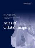
Atlas of Orbital Imaging
by Guy Ben Simon Gahl Greenberg Daphna Landau PratThis book features in-depth descriptions of imaging modalities (MRI, CT, US, PET) for all orbital pathologies, including tumors, vascular anomalies, congenital anomalies, trauma, inflammations and infections. It describes all the imaging features of the pathologies, and includes guidance for differential diagnosis and relevant clinical data. Atlas of Orbital Imaging serves as a clinical and educational resource for ophthalmologists/orbital surgeon residents, as well as a source of reference for consultants and neuroradiologists at all levels. The illustrations are both highly detailed and depict the orbit in vivid colour, adding to the attractiveness of the chapters. This reference work is a worldwide collaborative effort of all leading orbital surgeons and neuro-radiologists (in Europe, America, Australia and Asia) and provides an indispensable resource for developing skills and knowledge of orbital imaging.
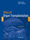
Atlas of Organ Transplantation
by Abhinav Humar Mark L. SturdevantAtlas of Organ Transplantation, Second Edition, provides the reader with a comprehensive and pictorial step-by-step account of abdominal organ transplant procedures performed by contemporary transplant surgeons today. Emphasis is placed on newer procedures or procedures that have undergone significant modifications. It is recognized that there are many well-accepted techniques for the same procedure, with each having potential merit. While it is impossible to present all of these variations, an attempt is made to describe the common variations in surgical technique and common variation in procedure based on anatomical variations. Written by an expert in the field, Atlas of Organ Transplantation, Second Edition, includes schematic diagrams and high-quality intraoperative photographs, allowing readers to clearly visualize the course of the operative procedure. This format provides the reader with a clear visual and written description of all major transplant procedures in one reference book.
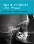
Atlas of Orthodontic Case Reviews
by Marjan Askari Stanley A. AlexanderAtlas of Orthodontic Case Reviews offers a comprehensive resource to the treatment of orthodontic malocclusions with a case-based approach. Discusses and illustrates the treatment of orthodontic malocclusions using actual clinical cases Presents more than 800 clinical photographs showing the stages of each treatment, to act as a visual reference Includes a description of each malocclusion, an explanation of the desired treatment outcomes, an account of the changes, and review questions for each case
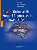
Atlas of Orthopaedic Surgical Approaches to the Lower Limbs
by Rosa Ballis Bujar H. Shabani Dafina BytyqiEnriched by original anatomical artworks this atlas comprehensively presents the diverse orthopaedic approaches to lower limb surgery. Combining clear visual information with a consistent and concise structure, including tips, tricks and pitfalls, this manual presents all the most used approaches in orthopaedic prosthetic surgery and traumatology. Ranging from the hip to the ankle each chapter includes a clear presentation of the joint’ s anatomy, with particular emphasis on vessels, nerves and other relevant anatomic structures. Beautiful water-colour illustrations realized by one of the authors accompany the reader step-by-step through each of the approaches described. This atlas offers a timely and up-to-date resource for both practicing and fellow orthopaedic surgeons with an interest in lower limb surgery.
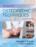
Atlas of Osteopathic Techniques
by Alexander S. Nicholas Evan A. NicholasEasy to navigate and rich with engaging learning features, the 4th edition of this bestselling, one-of-a-kind resource reflects the most up-to-date information on basic anatomical concepts and techniques to help users confidently comprehend and apply them.
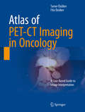
Atlas of PET-CT Imaging in Oncology: A Case-Based Guide to Image Interpretation
by Tamer Özülker Filiz ÖzülkerThis atlas is a case-based guide to the interpretation of FDG PET-CT images in clinical scenarios faced by physicians during the routine practice of oncology. The book aims to help the practitioner to overcome diagnostic dilemmas through familiarization with the physiologic distribution of FDG, normal variants and benign findings. The main focus, however, is the imaging of major oncological diseases. Different pathologies are addressed in individual chapters comprising teaching files of cases, each of which corresponds to a common indication for PET-CT imaging, such as metabolic characterization of lesions, staging, restaging and evaluation of response to therapy. Each case is accompanied by an explanation of the patient's history, interpretation of the PET-CT study, and a teaching point often supported by relevant literature. This book will be of great value to residents and practitioners in nuclear medicine, radiology, oncology, radiation oncology and nuclear medicine technology.
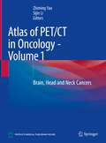
Atlas of PET/CT in Oncology - Volume 1: Brain, Head and Neck Cancers
by Zhiming Yao Sijin LiThe atlas aims to help practitioners to interpret PET/CT images of tumors in brain, head and neck in a timely and accurate way. Illustrating in a case-based manner, the PET/CT appearances of glioma, meningioma, nasopharyngeal carcinoma, eye tumors, ear and temporal bone tumors, and neck tumors are covered. Each chapter is organized in the same format of clinical overview, PET/CT diagnosis, typical/atypical/confusing manifestation of the diseases. This book will be a useful reference to residents and practitioners in nuclear medicine, radiology, oncology, radiation oncology and nuclear medicine technology.
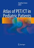
Atlas of PET/CT in Pediatric Patients
by Angelina CistaroThis richly illustrated book presents the pediatric applications of PET/CT in the full range of scenarios frequently encountered in a professional setting. It opens with a thorough introduction covering the fundamental science and the clinical basis for use of PET/CT in this age group. Pitfalls and artifacts are examined, and normal variations and benign findings are carefully described. Each subsequent chapter addresses the role of PET/CT with different radiopharmaceuticals in the evaluation and management of a specific disease. The full range of oncological diseases is covered, including the rare ones. Succinct descriptions of clinical cases are included and, when appropriate, comparisons are made with other modalities. In addition, the role of PET/CT in biopsy guidance and in radiation therapy planning is explained. This book will be invaluable for residents and practitioners in nuclear medicine, radiology, oncology, radiation oncology, and nuclear medicine technology