- Table View
- List View
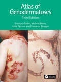
Atlas of Genodermatoses
by Gianluca Tadini Michela Brena Lidia Pezzani Francesca BesagniGenodermatoses are often considered rare diseases seldom seen by practicing clinicians, but as a result, professionals often have little experience or confidence with their diagnosis when they are called upon for a clinical case.This text presents a comprehensive illustrated overview of almost 200 inherited diseases of the skin, hair, and nails. Examples have been expanded, with new images added to provide clear examples, alongside coherent and comprehensive explanations to enable clinicians to easily identify and source relevant information. This resource encompasses a varied range of skin diseases, providing accessible and in-depth information to help familiarise clinicians. The entry for each disease provides background, followed by common characterisations, manifestations, laboratory findings, genetics, cutaneous and extracutaneous findings, differential diagnosis, an overview of complications and recommended follow-ups.Authored by dermatologists and geneticists, this is an atlas of scientific research which updates established information with current studies and references. In its third edition, this text becomes an invaluable resource for dermatologists and pediatricians.
Atlas of Geriatric Dermatology
by Robert A. Norman Edward M. Young JrThis is a comprehensive, practical, densely illustrated diagnostic and therapeutic guide for all geriatric dermatology providers. The book comprises 50 chapters and over 600 color photographs on topics ranging from common conditions such as basal cell carcinoma, rosacea, and seborrheic dermatitis to unusual conditions such as angiosarcoma, dermatofibrosarcoma protuberans, and porphyria cutanea tarda. Sections include: - Inflammatory conditions (including contact dermatitis, alopecia, erythema multiforme, pemphigus, bullous pemphigoid, porphyria, pruritus, psoriasis, rosacea, seborrhea, urticaria, xerosis, and more) - Infections (fungus, herpes simplex and zoster, scabies, lice, and warts) - Skin signs in systemic disease (skin tags, cutaneous metastases, xanthomas) - Regional dermatoses (intertrigo, leg ulcers, pressure sores) - Benign tumors (chondrodermatitis, cysts, ganglion, fibrous papule, seborrheic keratoses, lentigines, and benign vascular lesions) - Pre-malignant and malignant tumors (actinic keratoses, angiosarcoma, basal cell carcinoma, dermatofibroma and dermatofibrosarcoma protuberans, intraepidermal neoplasia, Kaposi's sarcoma, keratoacanthoma, lentigo maligna, cutaneous lymphoma, Mycosis fiungoides, melanoma, nevi and moles, and squamous cell carcinoma)
Atlas of Glaucoma
by Neil Choplin Carlo TraversoGlaucoma affects all age groups and is a leading cause of blindness worldwide. It is imperative that practicing clinicians and surgeons recognize both primary and secondary glaucoma as well as cases of glaucoma associated with other disorders. Atlas of Glaucoma, Third Edition provides an in-depth review and analysis of the management of glaucoma an
Atlas of Global Change Risk of Population and Economic Systems (IHDP/Future Earth-Integrated Risk Governance Project Series)
by Peijun ShiThis book is open access and illustrates the spatial distribution of the global change risk of population and economic systems with the maps of environment, global climate change, global population and economic systems, and global change risk. The risks of global change are mapped at 0.25 degree grid unit. The risk results and their contribution rates of the world at national level are unprecedentedly derived and ranked. The book can be a good reference for researchers and students in the field of global climate change and natural disaster risk management, as well as risk managers and enterpriser to understand the global change risk of population and economic systems.
Atlas of Global Development
by The World BankPublished in association with Harper Collins, the completely revised and updated third edition of the Atlas of Global Development vividly illustrates the key development challenges facing our world today. 'This is an excellent, up-to-date source book which will be invaluable for students of, and staff teaching, higher levels of geography .... a clear, concise, easily-accessible and well-illustrated volume.' - Geographical Association, United Kingdom
Atlas of Graft-versus-Host Disease: Approaches to Diagnosis and Treatment
by Jonathan A. CotliarThis atlas provides trainees and practicing physicians a visual toolkit to help recognize and manage this difficult condition appropriately. This text highlights the clinical variability of GVHD, and arms readers with the diagnostic clues to categorize patients according to the current grading/staging guidelines. Furthermore, this atlas offers evidence-based diagnostic and treatment algorithms for physicians to use while at patients' bedsides.
Atlas of Great Comets
by Ronald StoyanThroughout the ages, comets, enigmatic and beautiful wandering objects that appear for weeks or months, have alternately fascinated and terrified humankind. The result of five years of careful research, Atlas of Great Comets is a generously illustrated reference on thirty of the greatest comets that have been witnessed and documented since the Middle Ages. Special attention is given to the cultural and scientific impact of each appearance, supported by a wealth of images, from woodcuts, engravings, historical paintings and artifacts, to a showcase of the best astronomical photos and images. Following the introduction, giving the broad historical context and a modern scientific interpretation, the Great Comets feature in chronological order. For each, there is a contemporary description of its appearance along with its scientific, cultural and historical significance. Whether you are an armchair astronomer or a seasoned comet-chaser, this spectacular reference deserves a place on your shelf.
Atlas of Gynecologic Laparoscopy, Robotic-Assisted Laparoscopic Surgery, and Hysteroscopy: An Essential Surgical Guide
by Susan Khalil Konstantin ZakashanskyThis surgical atlas of laparoscopic, hysteroscopic, and robotic gynecologic procedures includes contemporary images. With endoscopic innovation and technology this resource includes access to high quality images essential for medical education and surgical procedures.This atlas serves to address gaps in high quality, up-to-date images and online access to a concise reference. This atlas includes surgical anatomy and different disease sites, including the uterus, ovaries, and vasculature of the pelvis. Chapters then move to cover specific diseases and the common and uncommon gynecologic procedures to treat them. This includes endometriosis, uterine pathology, and adnexal masses/ovarian cysts. Chapters are enhanced by high quality images and online reference access. This is an ideal guide for gynecological surgeons, gynecologic surgical subspecialty fellows, gynecologists, and residents.
Atlas of Gynecologic Oncology Imaging (Atlas of Oncology Imaging)
by Oguz AkinThis book provides a comprehensive visual review of pathologic disease variations of the five main types of gynecologic cancers: ovarian, endometrial, cervical, vaginal, and vulvar Through the use of cross-sectional imaging modalities, including computed tomography, magnetic resonance imaging, ultrasound, and positron emission tomography, it depicts normal anatomy as well as common gynecological tumors For each type of cancer, aspects such as primary staging, recurrence patterns, and findings from different yet complementary imaging modalities are explored. Atlas of Gynecologic Oncology Imaging presents a coherent perspective of the roles of standard and cutting-edge imaging techniques in gynecologic oncology via a multidisciplinary approach to cancer care. Featuring over 600 images, this book is a valuable resource for diagnostic radiologists, radiation oncologists, and gynecologists.
Atlas of Handheld Ultrasound
by Eric Topol Jagat Narula Bret P. Nelson Anjali Bhagra Sharon L. MulvaghThis atlas comprehensively covers the use of basic ultrasound (US) scanning technologies to recognize both normal and pathologic states. It covers how to use US for soft tissue diagnosis, assessing cardiac function, pericardial effusion, tamponade, peripheral veins, gallbladder, bowel, kidneys, skull, sinuses, eye, pleura, scrotum and testes. Atlas of Handheld Ultrasound provides detailed clinically relevant examples of how US can be used to aid diagnosis across a wide range of medical disciplines. Chapters are formatted in a clear and easy-to-follow format, with a range of interactive material to aid the reader in developing a deep understanding of how to use and interpret US correctly. It therefore provides a critical resource on how to use the modality for both specialist and non-specialist practitioners.
Atlas of Head Neck and Skull-base Surgery
by Rupa Mehta Nitin M Nagarkar Ambesh Singh Karthik N Rao Prajwal S DangeHead and neck anatomy is fairly constant, meticulous dissection of the structures and forms the backbone of a successful surgery. The entire diverse spectrum of head neck cancers, thyroid lesions, neural, lymphangiomatous lesions, and carotid body tumors are discussed along with preop, intraop, and postop follow-up pictures. The painstaking selection of intraoperative photographs of various patients operated at our center with an aim to demonstrate the finesse of the surgical management is the motive behind this atlas. The reader picking up this beautifully illustrated volume draws on the surgical expertise of an impressively well-trained surgical team. The atlas will be a pleasure to read and a powerful force to grab the advance treatment standards in the huge variety of head and neck disorders. This atlas will be a valuable resource to aspiring head and neck surgeons and oncologists, and it will act as a volume to refer to in the surgical management of head and neck disorders.
Atlas of Head and Neck Cancer Surgery: The Compartment Surgery for Resection in 3-D
by Nirav TrivediSurgery for head neck cancer has evolved greatly in the recent years. Appropriate surgical resection with negative margins still remain corner-stone in achieving good oncological outcome. This atlas proposes a new concept of " The Compartment Surgery " to achieve negative margins in third dimensions which is the problem area in majority of cases. Reconstructive techniques have evolved vastly in the recent years with use of microvascular free flaps and this has significantly improved the functional outcome. Performing each step in appropriate manner cumulatively enables us to perform more complex procedures. Theme of this atlas revolves around this fact. This atlas on head and neck cancers takes a fresh stylistic approach where each surgical procedure is described in a step-wise manner through labeled high-resolution images with minimal incorporation of text. This allows the surgeons to rapidly revise the operative steps of a procedure within minutes just before they start a surgery. Head and neck has multidimensional anatomy and surgical treatment requires specific approach for each subsite. This operative atlas covers the entire spectrum of common, uncommon and rare cancers of the head and neck area. Each subsite is addressed in a separate chapter with further subdivisions for surgery of tumors with varying extent. Each procedure is demonstrated with photographs of each surgical step and line diagrams. Commonly used flaps (regional and free flaps) are demonstrated in separate chapter for reconstruction. With over 1000 images, and coverage of both the ablative and reconstructive surgical procedures, this is a technique-focused atlas in comparison to the available comprehensive texts. There are two chapters on very advanced cancers, and surgery in resource constrained surgical units making the book relevant to a wide range of cancer surgeons and fellows-in-training in varied clinical settings.
Atlas of Head and Neck Endocrine Disorders: Special Focus on Imaging and Imaging-Guided Procedures
by Luca Giovanella Giorgio Treglia Roberto ValcaviThis atlas draws on a multidisciplinary approach to provide a comprehensive overview on endocrine disorders of the head and neck, with particular emphasis on the role of imaging and image-guided procedures. The first section discusses the basic characteristics of the imaging methods and other techniques used for evaluation and diagnosis. The remainder of the book focuses on application of these methods in thyroid, parathyroid, and other endocrine disorders of the head and neck. The coverage is wide ranging, encompassing Graves' disease, toxic multinodular goiter, toxic adenoma, thyroiditis, non-toxic goiter, benign nodules, and the different forms of thyroid carcinoma, as well as parathyroid adenoma, hyperplasia, and carcinoma and paragangliomas. Informative, high-quality images are provided by international experts in endocrine disorders, including endocrinologists, pathologists, radiologists, nuclear medicine physicians, and surgeons, who also discuss sample cases and provide syntheses of the relevant scientific literature.
Atlas of Head and Neck Robotic Surgery
by Ziv Gil Moran Amit Michael E. KupfermanThis atlas offers precise, step-by-step descriptions of robotic surgical techniques in the fields of otolaryngology and head and neck surgery, with the aim of providing surgeons with a comprehensive guide. The coverage encompasses all current indications and the full range of robotic surgical approaches, including transoral, transaxillary, transmaxillary, and transcervical. Key clinical and technical issues and important aspects of surgical anatomy are highlighted, and advice is provided on ancillary topics such as postoperative care and robotic reconstructive surgery. Robotic surgery has proved a significant addition to the armamentarium of tools in otolaryngology and head and neck surgery. It is now used in many centers as the workhorse for resection of oropharyngeal and laryngeal tumors, thyroid surgery, and base of tongue resection in patients with obstructive sleep apnea. The da Vinci robotic system, with its three-dimensional vision system, is also excellent for parapharyngeal, nasopharyngeal, and skull base resections. This superbly illustrated book, with accompanying online videos, will be ideal for residents in otolaryngology-head and neck surgery and skull base surgery who are working in a robotic cadaver lab and for specialists seeking to further improve their dissection techniques.
Atlas of Head and Neck Surgery (Springer Surgery Atlas Series)
by Rui Fernandes Ricard Simo Paul PracyThis atlas aims to provide the reader with comprehensive and structured knowledge of contemporary head and neck surgical procedures in patients with both benign and malignant diseases. The bulk of the atlas is devoted to the surgical management of malignant tumors of the upper aerodigestive tract, with a separate chapter focusing on each major anatomic subsite. All aspects of endoscopy are covered, as is surgery of the upper airway, including tracheostomy, laryngotracheal reconstruction and surgery for vocal cord paralysis. Thorough consideration is also given to procedures for the treatment of carotid body tumors, branchial arch anomalies, deep neck space infections, pharyngeal pouches, and benign disease of the thyroid, parathyroid, and salivary glands. A final chapter addresses in detail the reconstruction of surgical defects in the head and neck. Each chapter includes bespoke drawings and diagrams to illustrate specific technical points and surgical steps. The authors are leading head and neck surgeons from Europe, North America, India, Africa, and Australia. Readers seeking a better understanding of how to carry out surgical procedures in this anatomic region will find the atlas to be an invaluable aid.
Atlas of Head/Neck and Spine Normal Imaging Variants
by Alexander McKinney Zuzan Cayci Mehmet Gencturk David Nascene Matt Rischall Jeffrey Rykken Frederick OttThis text provides a comprehensive overview of the normal variations of the neck, spine, temporal bone and face that may simulate disease. Comprised of seven chapters, this atlas focuses on specific topical variations, among them head-neck variants, orbital variants, sinus, and temporal bone variants, and cervical, thoracic, and lumbar variations of the spine. It also includes comparison cases of diseases that should not be confused with normal variants. Atlas of Head/Neck and Spine Normal Imaging Variants is a much needed resource for a diverse audience, including neuroradiologists, neurosurgeons, neurologists, orthopedists, emergency room physicians, family practitioners, and ENT surgeons, as well as their trainees worldwide.
Atlas of Heart Anatomy and Development
by Florin Mihail FilipoiuThis heart anatomy book describes the cardiac development and cardiac anatomy in the development of the adult heart, and is illustrated by numerous images and examples. It contains 550 images of dissected embryo and adult hearts, obtained through the dissection and photography of 235 hearts. It has been designed to allow the rapid understanding of the key concepts and that everything should be clearly and graphically explained in one book. This is an atlas of cardiac development and anatomy of the human heart which distinguishes itself with the use of 550 images of embryonic, fetal and adult hearts and using text that is logical and concise. All the mentioned anatomical structures are shown with the use of suggestive dissection images to emphasize the details and the overall location. All the images have detailed comments, while clinical implications are suggested. The dissections of different hearts exemplify the variability of the cardiac structures. The electron and optical microscopy images are sharp and provide great fidelity. The arterial molds obtained using methyl methacrylate are illustrative and the pictures use suggestive angles. The dissections were made on human normal and pathological hearts of different ages, increasing the clinical utility of the material contained within.
Atlas of Hepatocellular Carcinoma Pathology
by Haeryoung Kim Wei-Qiang Leow Regina Lo Paulo Giovanni Mendoza Anthony Wing-Hung ChanThis atlas is designed to be a user-friendly bench-side reference for pathology trainees and general pathologists in handling and interpreting specimens of hepatocellular carcinoma. It provides over 550 high-quality gross and microscopic photos focusing on hepatocellular carcinoma and its mimickers, and demonstrating a full range of various histological variants of hepatocellular carcinoma. Introductory text in each chapter summarises salient clinical associations, pathological features, and molecular alterations of different variants of hepatocellular carcinoma. Differentiation between hepatocellular carcinoma and its mimickers is illustrated through various case studies.The authors are nationally and internationally recognized hepatopathologists in the Asian-Pacific regions (Hong Kong, Korea, the Philippines, and Singapore), in which the incidence of hepatocellular carcinoma is high.The authors are nationally and internationally recognized hepatopathologists in the Asian-Pacific regions (Hong Kong, Korea, the Philippines, and Singapore), in which the incidence of hepatocellular carcinoma is high.The authors are nationally and internationally recognized hepatopathologists in the Asian-Pacific regions (Hong Kong, Korea, the Philippines, and Singapore), in which the incidence of hepatocellular carcinoma is high.The authors are nationally and internationally recognized hepatopathologists in the Asian-Pacific regions (Hong Kong, Korea, the Philippines, and Singapore), in which the incidence of hepatocellular carcinoma is high.The authors are nationally and internationally recognized hepatopathologists in the Asian-Pacific regions (Hong Kong, Korea, the Philippines, and Singapore), in which the incidence of hepatocellular carcinoma is high.The authors are nationally and internationally recognized hepatopathologists in the Asian-Pacific regions (Hong Kong, Korea, the Philippines, and Singapore), in which the incidence of hepatocellular carcinoma is high.The authors are nationally and internationally recognized hepatopathologists in the Asian-Pacific regions (Hong Kong, Korea, the Philippines, and Singapore), in which the incidence of hepatocellular carcinoma is high.
Atlas of High-Resolution Manometry, Impedance, and pH Monitoring
by Mark Pimentel Sarvee Moosavi Ali Rezaie Nipaporn PichetshoteThis atlas provides a concise yet comprehensive overview of high-resolution manometry, impedance and pH monitoring. Through instructive text and over 130 high-yield images, the atlas describes the basic principles of esophageal, antroduodenal and anorectal high-resolution manometry, reviews both normal and pathologic findings on manometry, covers technical aspects of pH monitoring and impedance, and outlines advances in equipment, software, and diagnostic guidelines.Written by experts in the field, Atlas of High-Resolution Manometry, Impedance, and pH Monitoring is a valuable resource for gastroenterologists and other clinicians and practitioners who work or are interested in the GI motility field.
Atlas of Histology with Functional Correlations: With Functional Correlations (Point (lippincott Williams And Wilkins) Ser.)
by Victor P. EroschenkoThis thirteenth edition of Atlas of Histology with Functional Correlations (formerly diFiore’s) provides a rich understanding of the basic histology concepts that medical and allied health students need to know.
Atlas of Human Anatomy
by Frank H. NetterAtlas of Human Anatomy uses Frank H. Netter, MD's detailed illustrations to demystify this often intimidating subject, providing a coherent, lasting visual vocabulary for understanding anatomy and how it applies to medicine. This 5th Edition features a stronger clinical focus-with new diagnostic imaging examples-making it easier to correlate anatomy with practice. Student Consult online access includes supplementary learning resources, from additional illustrations to an anatomy dissection guide and more. Netter. It's how you know. See anatomy from a clinical perspective with hundreds of exquisite, hand-painted illustrations created by, and in the tradition of, pre-eminent medical illustrator Frank H. Netter, MD. Join the global community of healthcare professionals who've mastered anatomy the Netter way Expand your study at studentconsult. com, where you'll find a suite of learning aids including selected Netter illustrations, additional clinically-focused illustrations and radiologic images, videos from Netter's 3D Interactive Anatomy, dissection modules, an anatomy dissection guide, multiple-choice review questions, drag-and-drop exercises, clinical pearls, clinical cases, survival guides, surgical procedures, and more. Correlate anatomy with practice through an increased clinical focus, many new diagnostic imaging examples, and bonus clinical illustrations and guides online.
Atlas of Human Body Ultrasound Scanning: Methods and Diagnostic Applications
by Mei ZhangThis atlas describes the diagnosis practice and cases of human body ultrasound. It includes anatomic section, standard scan ultrasonogram of every organ, scanning methods and key points, measurement methods, normal value ranges, and clinical significances of every sections. Providing basic information and fundamental principles of ultrasonic diagnosis, it discusses ultrasound scanning of 14 organs in individual chapters. Each section sonography is accompanied by the scanning method, section structure, measuring method and clinical application. The uniform structure and detailed instructions make this atlas an easy-to-use resource for residents to refer to when they encounter specific ultrasound diagnostic problems.
Atlas of Human Hair: Microscopic Characteristics
by Robert R. Ogle Jr. Michelle J. FoxIt fills a void in the resources available to researchers and practitioners in forensic hair examination by providing photographic archetypes for the microscopic characteristics of human hair and the variates of the characteristics seen in forensic examinations, including curl; color; pigment distribution and density; cortical fusi; and ovoid bodie
Atlas of Human Infectious Diseases
by Peter Horby John P. Woodall Heiman F.L.WertheimThe Atlas of Human Infectious Diseases provides a much needed practical and visual overview of the current distribution and determinants of major infectious diseases of humans. The comprehensive full-color maps show at a glance the areas with reported infections and outbreaks, and are accompanied by a concise summary of key information on the infectious agent and its clinical and epidemiological characteristics. Since infectious diseases are dynamic, the maps are presented in the context of a changing world, and how these changes are influencing the geographical distribution on human infections. This unique atlas: Contains more than 145 high quality full-color maps covering all major human infectious diseases Provides key information on the illustrated infectious diseases Has been compiled and reviewed by an editorial board of infectious disease experts from around the world The result is a concise atlas with a consistent format throughout, where material essential for understanding the global spatial distribution of infectious diseases has been thoughtfully assembled by international experts. Atlas of Human Infectious Diseases is an essential tool for infectious disease specialists, medical microbiologists, virologists, travel medicine specialists, and public health professionals. The Atlas of Human Infectious Diseases is accompanied by a FREE enhanced Wiley Desktop Edition - an interactive digital version of the book with downloadable images and text, highlighting and note-taking facilities, book-marking, cross-referencing, in-text searching, and linking to references and glossary terms.
Atlas of Human Pluripotent Stem Cells in Culture
by Lyn Healy Ludmila RubanThis lavishly-illustrated, authoritative atlas explores the intricate art of culturing human pluripotent stem cells. Twelve chapters - containing more than 280 color illustrations - cover a variety of topics in pluripotent stem cell culturing including mouse and human fibroblasts, human embryonic stem cells and induced pluripotent stem cells, characteristic staining patterns, and abnormal cultures, among others. Atlas of Human Pluripotent Stem Cells in Culture is a comprehensive collection of illustrated techniques complemented by informative and educational captions examining what good quality cells look like and how they behave in various environments. Examples of perfect cultures are compared side-by-side to less-than-perfect and unacceptable examples of human embryonic and induced pluripotent stem cell colonies. This detailed and thorough atlas is an invaluable resource for researchers, teachers, and students who are interested in or working with stem cell culturing.
